 Movie
Movie Controller


+ Open data

- Basic information
| Entry | Database: PDB / ID: 5wxm | ||||||
|---|---|---|---|---|---|---|---|
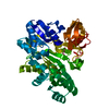
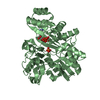
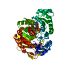
| Title | Crystal structure of the Imp3 and Mpp10 complex | ||||||
 Components Components |
| ||||||
 Keywords Keywords | RIBOSOMAL PROTEIN / protein complex / components of 90S preribosome | ||||||
| Function / homology |  Function and homology information Function and homology informationMpp10 complex / endonucleolytic cleavage in 5'-ETS of tricistronic rRNA transcript (SSU-rRNA, 5.8S rRNA, LSU-rRNA) / endonucleolytic cleavage to generate mature 5'-end of SSU-rRNA from (SSU-rRNA, 5.8S rRNA, LSU-rRNA) / RNA folding chaperone / sno(s)RNA-containing ribonucleoprotein complex / snoRNA binding / Major pathway of rRNA processing in the nucleolus and cytosol / 90S preribosome / endonucleolytic cleavage in ITS1 to separate SSU-rRNA from 5.8S rRNA and LSU-rRNA from tricistronic rRNA transcript (SSU-rRNA, 5.8S rRNA, LSU-rRNA) / maturation of SSU-rRNA ...Mpp10 complex / endonucleolytic cleavage in 5'-ETS of tricistronic rRNA transcript (SSU-rRNA, 5.8S rRNA, LSU-rRNA) / endonucleolytic cleavage to generate mature 5'-end of SSU-rRNA from (SSU-rRNA, 5.8S rRNA, LSU-rRNA) / RNA folding chaperone / sno(s)RNA-containing ribonucleoprotein complex / snoRNA binding / Major pathway of rRNA processing in the nucleolus and cytosol / 90S preribosome / endonucleolytic cleavage in ITS1 to separate SSU-rRNA from 5.8S rRNA and LSU-rRNA from tricistronic rRNA transcript (SSU-rRNA, 5.8S rRNA, LSU-rRNA) / maturation of SSU-rRNA / small-subunit processome / rRNA processing / ribosomal small subunit biogenesis / rRNA binding / nucleolus / nucleoplasm / identical protein binding / nucleus Similarity search - Function | ||||||
| Biological species |  | ||||||
| Method |  X-RAY DIFFRACTION / SYNCHROTRON / SIRAS / Resolution: 2.304 Å X-RAY DIFFRACTION / SYNCHROTRON / SIRAS / Resolution: 2.304 Å | ||||||
 Authors Authors | Ye, K. / Zheng, S. | ||||||
 Citation Citation | Journal: Elife / Year: 2017 Title: Molecular architecture of the 90S small subunit pre-ribosome. Authors: Qi Sun / Xing Zhu / Jia Qi / Weidong An / Pengfei Lan / Dan Tan / Rongchang Chen / Bing Wang / Sanduo Zheng / Cheng Zhang / Xining Chen / Wei Zhang / Jing Chen / Meng-Qiu Dong / Keqiong Ye /  Abstract: Eukaryotic small ribosomal subunits are first assembled into 90S pre-ribosomes. The complete 90S is a gigantic complex with a molecular mass of approximately five megadaltons. Here, we report the ...Eukaryotic small ribosomal subunits are first assembled into 90S pre-ribosomes. The complete 90S is a gigantic complex with a molecular mass of approximately five megadaltons. Here, we report the nearly complete architecture of 90S determined from three cryo-electron microscopy single particle reconstructions at 4.5 to 8.7 angstrom resolution. The majority of the density maps were modeled and assigned to specific RNA and protein components. The nascent ribosome is assembled into isolated native-like substructures that are stabilized by abundant assembly factors. The 5' external transcribed spacer and U3 snoRNA nucleate a large subcomplex that scaffolds the nascent ribosome. U3 binds four sites of pre-rRNA, including a novel site on helix 27 but not the 3' side of the central pseudoknot, and crucially organizes the 90S structure. The 90S model provides significant insight into the principle of small subunit assembly and the function of assembly factors. | ||||||
| History |
|
- Structure visualization
| Structure viewer | Molecule: MolmilJmol/JSmol |
|---|
- Downloads & links
-Download
| PDBx/mmCIF format | 5wxm.cif.gz | 84.9 KB | Display | PDBx/mmCIF format |
|---|---|---|---|---|
| PDB format | pdb5wxm.ent.gz | 63.7 KB | Display | PDB format |
| PDBx/mmJSON format | 5wxm.json.gz | Tree view | PDBx/mmJSON format | |
| Others |  Other downloads Other downloads |
-Validation report
| Arichive directory | https://data.pdbj.org/pub/pdb/validation_reports/wx/5wxmftp://data.pdbj.org/pub/pdb/validation_reports/wx/5wxm | HTTPS FTP |
|---|
-Related structure data
| Related structure data | 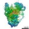 6695C 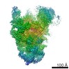 6696C  6697C 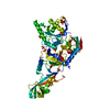 5wwnC 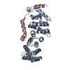 5wwoC 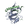 5wxlC 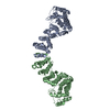 5wy3C  5wyjC  5wykC 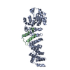 5wylC C: citing same article ( |
|---|---|
| Similar structure data |


-Links
 PDBj
PDBj

- Assembly
| Deposited unit | 
| ||||||||
|---|---|---|---|---|---|---|---|---|---|
| 1 |
| ||||||||
| Unit cell |
|
-Components
| #1: Protein | Mass: 19053.246 Da / Num. of mol.: 2 / Fragment: UNP residues 26-183 Source method: isolated from a genetically manipulated source Source: (gene. exp.)  #2: Protein/peptide | Mass: 3756.327 Da / Num. of mol.: 2 / Fragment: UNP residues 430-461 Source method: isolated from a genetically manipulated source Source: (gene. exp.) #3: Chemical | ChemComp-SO4 /   Mass: 96.063 Da / Num. of mol.: 5 / Source method: obtained synthetically / Formula: SO4 Mass: 96.063 Da / Num. of mol.: 5 / Source method: obtained synthetically / Formula: SO4#4: Water | ChemComp-HOH / |  Mass: 18.015 Da / Num. of mol.: 192 / Source method: isolated from a natural source / Formula: H2O Mass: 18.015 Da / Num. of mol.: 192 / Source method: isolated from a natural source / Formula: H2OHas protein modification | Y | |
|---|
-Experimental details
-Experiment
| Experiment | Method: X-RAY DIFFRACTION / Number of used crystals: 1 |
|---|
- Sample preparation
| Crystal | Density Matthews: 2.18 Å3/Da / Density % sol: 43.58 % |
|---|---|
| Crystal grow | Temperature: 293 K / Method: vapor diffusion, hanging drop / pH: 5 Details: 0.2 M potassium sodium tartrate, 0.1 M sodium citrate, 1.7 M ammonium sulfate |
-Data collection
| Diffraction | Mean temperature: 100 K |
|---|---|
| Diffraction source | Source: SYNCHROTRON / Site: SSRF / Beamline: BL17U / Wavelength: 0.97915 Å |
| Detector | Type: ADSC QUANTUM 315r / Detector: CCD / Date: Jan 1, 2013 |
| Radiation | Protocol: SINGLE WAVELENGTH / Monochromatic (M) / Laue (L): M / Scattering type: x-ray |
| Radiation wavelength | Wavelength: 0.97915 Å / Relative weight: 1 |
| Reflection | Resolution: 2.3→50 Å / Num. obs: 18226 / % possible obs: 99.9 % / Redundancy: 6.2 % / Net I/σ(I): 15.4 |
- Processing
| Software |
| |||||||||||||||||||||||||||||||||||||||||||||||||
|---|---|---|---|---|---|---|---|---|---|---|---|---|---|---|---|---|---|---|---|---|---|---|---|---|---|---|---|---|---|---|---|---|---|---|---|---|---|---|---|---|---|---|---|---|---|---|---|---|---|---|
| Refinement | Method to determine structure: SIRAS / Resolution: 2.304→33.629 Å / SU ML: 0.24 / Cross valid method: FREE R-VALUE / σ(F): 1.35 / Phase error: 24.34 / Stereochemistry target values: ML
| |||||||||||||||||||||||||||||||||||||||||||||||||
| Solvent computation | Shrinkage radii: 0.9 Å / VDW probe radii: 1.11 Å / Solvent model: FLAT BULK SOLVENT MODEL | |||||||||||||||||||||||||||||||||||||||||||||||||
| Displacement parameters | Biso max: 84.87 Å2 / Biso mean: 26.6306 Å2 / Biso min: 7.97 Å2 | |||||||||||||||||||||||||||||||||||||||||||||||||
| Refinement step | Cycle: final / Resolution: 2.304→33.629 Å
| |||||||||||||||||||||||||||||||||||||||||||||||||
| Refine LS restraints |
| |||||||||||||||||||||||||||||||||||||||||||||||||
| LS refinement shell | Refine-ID: X-RAY DIFFRACTION / Total num. of bins used: 6
|
