 Movie
Movie Controller


+ Open data

- Basic information
| Entry | Database: PDB / ID: 7kv1 | ||||||
|---|---|---|---|---|---|---|---|
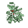
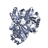
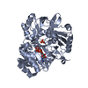
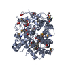
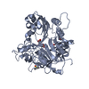
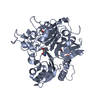
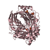
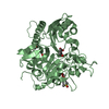
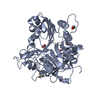
| Title | Surface glycan-binding protein A from Bacteroides uniformis | ||||||
 Components Components | SusD family protein | ||||||
 Keywords Keywords | SUGAR BINDING PROTEIN / SusD / Tetratricopeptide repeat / CBM | ||||||
| Function / homology | SusD-like, N-terminal / Starch-binding associating with outer membrane / RagB/SusD domain / SusD family / Prokaryotic membrane lipoprotein lipid attachment site profile. / Tetratricopeptide-like helical domain superfamily / membrane / COBALT HEXAMMINE(III) / SusD family protein Function and homology information Function and homology information | ||||||
| Biological species |  Bacteroides uniformis (bacteria) Bacteroides uniformis (bacteria) | ||||||
| Method |  X-RAY DIFFRACTION / SYNCHROTRON / SAD / Resolution: 1.86 Å X-RAY DIFFRACTION / SYNCHROTRON / SAD / Resolution: 1.86 Å | ||||||
 Authors Authors | Tamura, K. / Brumer, H. / Van Petegem, F. | ||||||
| Funding support |  Canada, 1items Canada, 1items
| ||||||
 Citation Citation | Journal: J.Biol.Chem. / Year: 2021 Title: Distinct protein architectures mediate species-specific beta-glucan binding and metabolism in the human gut microbiota. Authors: Tamura, K. / Dejean, G. / Van Petegem, F. / Brumer, H. | ||||||
| History |
|
- Structure visualization
| Structure viewer | Molecule: MolmilJmol/JSmol |
|---|
- Downloads & links
-Download
| PDBx/mmCIF format | 7kv1.cif.gz | 205.9 KB | Display | PDBx/mmCIF format |
|---|---|---|---|---|
| PDB format | pdb7kv1.ent.gz | 161.3 KB | Display | PDB format |
| PDBx/mmJSON format | 7kv1.json.gz | Tree view | PDBx/mmJSON format | |
| Others |  Other downloads Other downloads |
-Validation report
| Arichive directory | https://data.pdbj.org/pub/pdb/validation_reports/kv/7kv1ftp://data.pdbj.org/pub/pdb/validation_reports/kv/7kv1 | HTTPS FTP |
|---|
-Related structure data
| Related structure data | 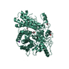 7kv2C 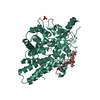 7kv3C 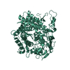 7kv4C  7kv5C 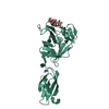 7kv6C  7kv7C 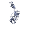 7kwbC 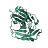 7kwcC C: citing same article ( |
|---|---|
| Similar structure data |

-Links
 PDBj
PDBj


- Assembly
| Deposited unit | 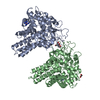
| ||||||||
|---|---|---|---|---|---|---|---|---|---|
| 1 | 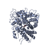
| ||||||||
| 2 | 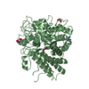
| ||||||||
| Unit cell |
|
-Components
| #1: Protein | Mass: 60535.145 Da / Num. of mol.: 2 Source method: isolated from a genetically manipulated source Source: (gene. exp.) Bacteroides uniformis (strain ATCC 8492 / DSM 6597 / CIP 103695 / JCM 5828 / NCTC 13054 / VPI 0061) (bacteria)Strain: ATCC 8492 / DSM 6597 / CIP 103695 / JCM 5828 / NCTC 13054 / VPI 0061 Gene: BACUNI_01488 / Production host: #2: Chemical |   Mass: 161.116 Da / Num. of mol.: 2 / Source method: obtained synthetically / Formula: CoH18N6 Mass: 161.116 Da / Num. of mol.: 2 / Source method: obtained synthetically / Formula: CoH18N6#3: Chemical |   Mass: 62.068 Da / Num. of mol.: 3 / Source method: obtained synthetically / Formula: C2H6O2 Mass: 62.068 Da / Num. of mol.: 3 / Source method: obtained synthetically / Formula: C2H6O2#4: Chemical | ChemComp-BTB / |   Mass: 209.240 Da / Num. of mol.: 1 / Source method: obtained synthetically / Formula: C8H19NO5 / Comment: pH buffer*YM Mass: 209.240 Da / Num. of mol.: 1 / Source method: obtained synthetically / Formula: C8H19NO5 / Comment: pH buffer*YM#5: Water | ChemComp-HOH / |  Mass: 18.015 Da / Num. of mol.: 550 / Source method: isolated from a natural source / Formula: H2O Mass: 18.015 Da / Num. of mol.: 550 / Source method: isolated from a natural source / Formula: H2OHas ligand of interest | N | |
|---|
-Experimental details
-Experiment
| Experiment | Method: X-RAY DIFFRACTION / Number of used crystals: 1 |
|---|
- Sample preparation
| Crystal | Density Matthews: 2.14 Å3/Da / Density % sol: 42.47 % |
|---|---|
| Crystal grow | Temperature: 295 K / Method: vapor diffusion Details: 0.1 M bis-tris pH 5.3, 0.2 M ammonium acetate, 22 % (w/v) PEG3350, 0.01 M hexamine cobalt (III) chloride |
-Data collection
| Diffraction | Mean temperature: 100 K / Serial crystal experiment: N |
|---|---|
| Diffraction source | Source: SYNCHROTRON / Site: APS  / Beamline: 23-ID-D / Wavelength: 1.0332 Å / Beamline: 23-ID-D / Wavelength: 1.0332 Å |
| Detector | Type: DECTRIS PILATUS3 6M / Detector: PIXEL / Date: Nov 18, 2020 |
| Radiation | Protocol: SINGLE WAVELENGTH / Monochromatic (M) / Laue (L): M / Scattering type: x-ray |
| Radiation wavelength | Wavelength: 1.0332 Å / Relative weight: 1 |
| Reflection | Resolution: 1.86→41.81 Å / Num. obs: 77609 / % possible obs: 99.9 % / Redundancy: 6.5 % / CC1/2: 0.996 / Net I/σ(I): 10.3 |
| Reflection shell | Resolution: 1.86→1.89 Å / Num. unique obs: 3967 / CC1/2: 0.578 |
- Processing
| Software |
| ||||||||||||||||||||||||||||||||||||||||||||||||||||||||||||
|---|---|---|---|---|---|---|---|---|---|---|---|---|---|---|---|---|---|---|---|---|---|---|---|---|---|---|---|---|---|---|---|---|---|---|---|---|---|---|---|---|---|---|---|---|---|---|---|---|---|---|---|---|---|---|---|---|---|---|---|---|---|
| Refinement | Method to determine structure: SAD / Resolution: 1.86→41.81 Å / Cor.coef. Fo:Fc: 0.964 / Cor.coef. Fo:Fc free: 0.948 / SU B: 3.263 / SU ML: 0.093 / Cross valid method: THROUGHOUT / σ(F): 0 / ESU R: 0.142 / ESU R Free: 0.134 / Stereochemistry target values: MAXIMUM LIKELIHOOD Details: HYDROGENS HAVE BEEN ADDED IN THE RIDING POSITIONS U VALUES : REFINED INDIVIDUALLY
| ||||||||||||||||||||||||||||||||||||||||||||||||||||||||||||
| Solvent computation | Ion probe radii: 0.8 Å / Shrinkage radii: 0.8 Å / VDW probe radii: 1.2 Å / Solvent model: MASK | ||||||||||||||||||||||||||||||||||||||||||||||||||||||||||||
| Displacement parameters | Biso max: 92.57 Å2 / Biso mean: 26.494 Å2 / Biso min: 13.56 Å2
| ||||||||||||||||||||||||||||||||||||||||||||||||||||||||||||
| Refinement step | Cycle: final / Resolution: 1.86→41.81 Å
| ||||||||||||||||||||||||||||||||||||||||||||||||||||||||||||
| Refine LS restraints |
| ||||||||||||||||||||||||||||||||||||||||||||||||||||||||||||
| LS refinement shell | Resolution: 1.86→1.903 Å / Rfactor Rfree error: 0
|
