 Movie
Movie Controller

[English] 日本語
 Yorodumi
- PDB-7kwc: Surface glycan-binding protein B (truncated) from Bacteroides the... -
+ Open data

- Basic information
| Entry | Database: PDB / ID: 7kwc | ||||||
|---|---|---|---|---|---|---|---|
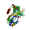
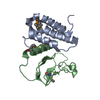
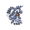
| Title | Surface glycan-binding protein B (truncated) from Bacteroides thetaiotaomicron | ||||||
 Components Components | BtSGBP-B_31-285 | ||||||
 Keywords Keywords | SUGAR BINDING PROTEIN / CBM / lectin | ||||||
| Function / homology | PKD domain superfamily / Galactose-binding-like domain superfamily / DUF5017 domain-containing protein Function and homology information Function and homology information | ||||||
| Biological species |  Bacteroides thetaiotaomicron (bacteria) Bacteroides thetaiotaomicron (bacteria) | ||||||
| Method |  X-RAY DIFFRACTION / SYNCHROTRON / SAD / Resolution: 2.611 Å X-RAY DIFFRACTION / SYNCHROTRON / SAD / Resolution: 2.611 Å | ||||||
 Authors Authors | Tamura, K. / Brumer, H. / Van Petegem, F. | ||||||
| Funding support |  Canada, 1items Canada, 1items
| ||||||
 Citation Citation | Journal: J.Biol.Chem. / Year: 2021 Title: Distinct protein architectures mediate species-specific beta-glucan binding and metabolism in the human gut microbiota. Authors: Tamura, K. / Dejean, G. / Van Petegem, F. / Brumer, H. | ||||||
| History |
|
- Structure visualization
| Structure viewer | Molecule: MolmilJmol/JSmol |
|---|
- Downloads & links
-Download
| PDBx/mmCIF format | 7kwc.cif.gz | 96.2 KB | Display | PDBx/mmCIF format |
|---|---|---|---|---|
| PDB format | pdb7kwc.ent.gz | Display | PDB format | |
| PDBx/mmJSON format | 7kwc.json.gz | Tree view | PDBx/mmJSON format | |
| Others |  Other downloads Other downloads |
-Validation report
| Arichive directory | https://data.pdbj.org/pub/pdb/validation_reports/kw/7kwcftp://data.pdbj.org/pub/pdb/validation_reports/kw/7kwc | HTTPS FTP |
|---|
-Related structure data
| Related structure data |  7kv1C 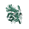 7kv2C 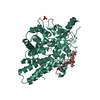 7kv3C 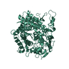 7kv4C  7kv5C 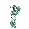 7kv6C  7kv7C  7kwbC C: citing same article ( |
|---|---|
| Similar structure data |


-Links
 PDBj
PDBj- Assembly
| Deposited unit | 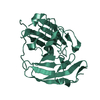
| ||||||||
|---|---|---|---|---|---|---|---|---|---|
| 1 |
| ||||||||
| Unit cell |
|
-Components
| #1: Protein | Mass: 31663.539 Da / Num. of mol.: 1 Source method: isolated from a genetically manipulated source Source: (gene. exp.) Bacteroides thetaiotaomicron (bacteria)Gene: HMPREF2534_00387 / Production host: |
|---|---|
| #2: Water | ChemComp-HOH /  Mass: 18.015 Da / Num. of mol.: 12 / Source method: isolated from a natural source / Formula: H2O Mass: 18.015 Da / Num. of mol.: 12 / Source method: isolated from a natural source / Formula: H2O |
-Experimental details
-Experiment
| Experiment | Method: X-RAY DIFFRACTION / Number of used crystals: 1 |
|---|
- Sample preparation
| Crystal | Density Matthews: 2.21 Å3/Da / Density % sol: 44.37 % |
|---|---|
| Crystal grow | Temperature: 295 K / Method: vapor diffusion Details: 0.1M magnesium chloride, 0.1M sodium citrate, 15% (w/v) PEG4000 |
-Data collection
| Diffraction | Mean temperature: 100 K / Serial crystal experiment: N |
|---|---|
| Diffraction source | Source: SYNCHROTRON / Site: SSRL  / Beamline: BL12-2 / Wavelength: 0.97946 Å / Beamline: BL12-2 / Wavelength: 0.97946 Å |
| Detector | Type: DECTRIS PILATUS3 6M / Detector: PIXEL / Date: Nov 30, 2020 |
| Radiation | Protocol: SINGLE WAVELENGTH / Monochromatic (M) / Laue (L): M / Scattering type: x-ray |
| Radiation wavelength | Wavelength: 0.97946 Å / Relative weight: 1 |
| Reflection | Resolution: 2.61→19.64 Å / Num. obs: 8556 / % possible obs: 94.5 % / Redundancy: 25.4 % / CC1/2: 1 / Net I/σ(I): 42.5 |
| Reflection shell | Resolution: 2.61→2.66 Å / Num. unique obs: 416 / CC1/2: 0.951 |
- Processing
| Software |
| |||||||||||||||||||||||||||||||||||||||||||||||||||||||||||||||||||||||||||||||||||||||||||||||||||||||||||||||||||||||||||||||||||||||||||||||||||||||||||
|---|---|---|---|---|---|---|---|---|---|---|---|---|---|---|---|---|---|---|---|---|---|---|---|---|---|---|---|---|---|---|---|---|---|---|---|---|---|---|---|---|---|---|---|---|---|---|---|---|---|---|---|---|---|---|---|---|---|---|---|---|---|---|---|---|---|---|---|---|---|---|---|---|---|---|---|---|---|---|---|---|---|---|---|---|---|---|---|---|---|---|---|---|---|---|---|---|---|---|---|---|---|---|---|---|---|---|---|---|---|---|---|---|---|---|---|---|---|---|---|---|---|---|---|---|---|---|---|---|---|---|---|---|---|---|---|---|---|---|---|---|---|---|---|---|---|---|---|---|---|---|---|---|---|---|---|---|
| Refinement | Method to determine structure: SAD / Resolution: 2.611→19.64 Å / Cor.coef. Fo:Fc: 0.928 / Cor.coef. Fo:Fc free: 0.902 / Cross valid method: FREE R-VALUE / ESU R: 1.15 / ESU R Free: 0.38 Details: Hydrogens have been added in their riding positions
| |||||||||||||||||||||||||||||||||||||||||||||||||||||||||||||||||||||||||||||||||||||||||||||||||||||||||||||||||||||||||||||||||||||||||||||||||||||||||||
| Solvent computation | Ion probe radii: 0.8 Å / Shrinkage radii: 0.8 Å / VDW probe radii: 1.2 Å / Solvent model: MASK BULK SOLVENT | |||||||||||||||||||||||||||||||||||||||||||||||||||||||||||||||||||||||||||||||||||||||||||||||||||||||||||||||||||||||||||||||||||||||||||||||||||||||||||
| Displacement parameters | Biso mean: 53.316 Å2
| |||||||||||||||||||||||||||||||||||||||||||||||||||||||||||||||||||||||||||||||||||||||||||||||||||||||||||||||||||||||||||||||||||||||||||||||||||||||||||
| Refinement step | Cycle: LAST / Resolution: 2.611→19.64 Å
| |||||||||||||||||||||||||||||||||||||||||||||||||||||||||||||||||||||||||||||||||||||||||||||||||||||||||||||||||||||||||||||||||||||||||||||||||||||||||||
| Refine LS restraints |
| |||||||||||||||||||||||||||||||||||||||||||||||||||||||||||||||||||||||||||||||||||||||||||||||||||||||||||||||||||||||||||||||||||||||||||||||||||||||||||
| LS refinement shell |
|
