[English] 日本語
Yorodumi
- PDB-5wsq: Crystal structure of C-Hg-T pair containing DNA duplex -

+
Open data


ID or keywords:

Loading...

-
Basic information

Entry
Database: PDB / ID: 5wsq
TitleCrystal structure of C-Hg-T pair containing DNA duplex
ComponentsDNA (5'-D(*GP*CP*CP*CP*GP*TP*GP*C)-3')
KeywordsDNA / DNA duplex / C-Hg-T pair
Function / homology: / 1,3-PROPANDIOL / DNA
Function and homology information
Biological speciesunidentified (others)
MethodX-RAY DIFFRACTION / SYNCHROTRON / SAD / Resolution: 1.05 Å
AuthorsGan, J.H. / Liu, H.H.
Funding support China, 2items
OrganizationGrant numberCountry
the Key Research and Development Project of China2016YFA0500600 China
National Natural Science Foundation of China31370728 China
CitationJournal: Nucleic Acids Res. / Year: 2017
Title: Flexibility and stabilization of HgII-mediated C:T and T:T base pairs in DNA duplex
Authors: Liu, H.H. / Cai, C. / Haruehanroengra, P. / Yao, Q.Q. / Chen, Y.Q. / Yang, C. / Luo, Q. / Wu, B.X. / Li, J.X. / Ma, J.B. / Sheng, J. / Gan, J.H.
History
DepositionDec 8, 2016Deposition site: PDBJ / Processing site: PDBJ
Revision 1.0Feb 8, 2017Provider: repository / Type: Initial release
Revision 1.1May 3, 2017Group: Database references
Revision 1.2Aug 30, 2017Group: Data collection / Category: diffrn_detector / Item: _diffrn_detector.detector
Revision 1.3Mar 20, 2024Group: Data collection / Database references / Derived calculations
Category: chem_comp_atom / chem_comp_bond ...chem_comp_atom / chem_comp_bond / database_2 / struct_conn
Item: _database_2.pdbx_DOI / _database_2.pdbx_database_accession ..._database_2.pdbx_DOI / _database_2.pdbx_database_accession / _struct_conn.ptnr1_auth_asym_id / _struct_conn.ptnr1_auth_comp_id / _struct_conn.ptnr1_auth_seq_id / _struct_conn.ptnr1_label_asym_id / _struct_conn.ptnr1_label_atom_id / _struct_conn.ptnr1_label_comp_id / _struct_conn.ptnr1_label_seq_id / _struct_conn.ptnr2_auth_asym_id / _struct_conn.ptnr2_auth_comp_id / _struct_conn.ptnr2_auth_seq_id / _struct_conn.ptnr2_label_asym_id / _struct_conn.ptnr2_label_atom_id / _struct_conn.ptnr2_label_comp_id / _struct_conn.ptnr2_label_seq_id

-
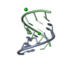
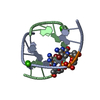
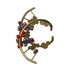
Structure visualization

Structure viewerMolecule:
MolmilJmol/JSmol

Downloads & links

-
Assembly

Deposited unit
A: DNA (5'-D(*GP*CP*CP*CP*GP*TP*GP*C)-3')
B: DNA (5'-D(*GP*CP*CP*CP*GP*TP*GP*C)-3')
hetero molecules


Theoretical massNumber of molelcules
Total (without water)5,2845
Polymers4,8072
Non-polymers4773
Water1,11762
1


  • Idetical with deposited unit
  • defined by author&software
TypeNameSymmetry operationNumber
identity operation1_555x,y,z1
Buried area1420 Å2
ΔGint-61 kcal/mol
Surface area2840 Å2
MethodPISA
Unit cell
Length a, b, c (Å)37.848, 51.138, 21.593
Angle α, β, γ (deg.)90.00, 90.00, 90.00
Int Tables number18
Space group name H-MP21212

-
Components

#1: DNA chain DNA (5'-D(*GP*CP*CP*CP*GP*TP*GP*C)-3')


Mass: 2403.580 Da / Num. of mol.: 2 / Source method: obtained synthetically / Source: (synth.) unidentified (others)
#2: Chemical ChemComp-HG / MERCURY (II) ION / Mercury (element)


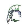
Mass: 200.590 Da / Num. of mol.: 2 / Source method: obtained synthetically / Formula: Hg
#3: Chemical ChemComp-PDO / 1,3-PROPANDIOL / 1,3-Propanediol


Mass: 76.094 Da / Num. of mol.: 1 / Source method: obtained synthetically / Formula: C3H8O2
#4: Water ChemComp-HOH / water / Water


Mass: 18.015 Da / Num. of mol.: 62 / Source method: isolated from a natural source / Formula: H2O

-
Experimental details

-
Experiment

ExperimentMethod: X-RAY DIFFRACTION / Number of used crystals: 1

-
Sample preparation

CrystalDensity Matthews: 2.17 Å3/Da / Density % sol: 43.41 %
Crystal growTemperature: 298 K / Method: vapor diffusion, hanging drop / Details: 10% v/v MPD

-
Data collection

DiffractionMean temperature: 100 K
Diffraction sourceSource: SYNCHROTRON / Site: SSRF / Beamline: BL19U1 / Wavelength: 0.979 Å
DetectorType: DECTRIS PILATUS3 S 6M / Detector: PIXEL / Date: Jun 30, 2016
RadiationProtocol: SINGLE WAVELENGTH / Monochromatic (M) / Laue (L): M / Scattering type: x-ray
Radiation wavelengthWavelength: 0.979 Å / Relative weight: 1
ReflectionResolution: 1.05→30 Å / Num. obs: 19098 / % possible obs: 98.1 % / Redundancy: 2 % / Rsym value: 0.12 / Net I/σ(I): 24
Reflection shellResolution: 1.05→1.09 Å / Redundancy: 1.9 % / CC1/2: 0.653 / % possible all: 96.4

-
Processing

Software
NameVersionClassification
REFMAC5.8.0135refinement
HKL-3000data reduction
HKL-3000data scaling
SHELXDEphasing
RefinementMethod to determine structure: SAD / Resolution: 1.05→30 Å / Cor.coef. Fo:Fc: 0.968 / Cor.coef. Fo:Fc free: 0.959 / Cross valid method: THROUGHOUT / ESU R: 0.026 / ESU R Free: 0.027 / Details: HYDROGENS HAVE BEEN ADDED IN THE RIDING POSITIONS
RfactorNum. reflection% reflectionSelection details
Rfree0.17491 1023 5.1 %RANDOM
Rwork0.15521 ---
obs0.1562 19098 99.45 %-
Solvent computationIon probe radii: 0.8 Å / Shrinkage radii: 0.8 Å / VDW probe radii: 1.2 Å
Displacement parametersBiso mean: 13.289 Å2
Baniso -1Baniso -2Baniso -3
1-0.12 Å2-0 Å20 Å2
2--0.01 Å2-0 Å2
3----0.14 Å2
Refinement stepCycle: 1 / Resolution: 1.05→30 Å
ProteinNucleic acidLigandSolventTotal
Num. atoms0 318 7 62 387
Refine LS restraints
Refine-IDTypeDev idealDev ideal targetNumber
X-RAY DIFFRACTIONr_bond_refined_d0.0090.011358
X-RAY DIFFRACTIONr_bond_other_d0.0010.02184
X-RAY DIFFRACTIONr_angle_refined_deg1.0971.172545
X-RAY DIFFRACTIONr_angle_other_deg2.0623432
X-RAY DIFFRACTIONr_dihedral_angle_1_deg
X-RAY DIFFRACTIONr_dihedral_angle_2_deg
X-RAY DIFFRACTIONr_dihedral_angle_3_deg
X-RAY DIFFRACTIONr_dihedral_angle_4_deg
X-RAY DIFFRACTIONr_chiral_restr0.0710.248
X-RAY DIFFRACTIONr_gen_planes_refined0.0110.02188
X-RAY DIFFRACTIONr_gen_planes_other0.0130.0284
X-RAY DIFFRACTIONr_nbd_refined
X-RAY DIFFRACTIONr_nbd_other
X-RAY DIFFRACTIONr_nbtor_refined
X-RAY DIFFRACTIONr_nbtor_other
X-RAY DIFFRACTIONr_xyhbond_nbd_refined
X-RAY DIFFRACTIONr_xyhbond_nbd_other
X-RAY DIFFRACTIONr_metal_ion_refined
X-RAY DIFFRACTIONr_metal_ion_other
X-RAY DIFFRACTIONr_symmetry_vdw_refined
X-RAY DIFFRACTIONr_symmetry_vdw_other
X-RAY DIFFRACTIONr_symmetry_hbond_refined
X-RAY DIFFRACTIONr_symmetry_hbond_other
X-RAY DIFFRACTIONr_symmetry_metal_ion_refined
X-RAY DIFFRACTIONr_symmetry_metal_ion_other
X-RAY DIFFRACTIONr_mcbond_it
X-RAY DIFFRACTIONr_mcbond_other
X-RAY DIFFRACTIONr_mcangle_it
X-RAY DIFFRACTIONr_mcangle_other
X-RAY DIFFRACTIONr_scbond_it0.6671.28357
X-RAY DIFFRACTIONr_scbond_other0.6641.279356
X-RAY DIFFRACTIONr_scangle_it
X-RAY DIFFRACTIONr_scangle_other0.8911.93546
X-RAY DIFFRACTIONr_long_range_B_refined1.23713.242534
X-RAY DIFFRACTIONr_long_range_B_other1.23613.244535
X-RAY DIFFRACTIONr_rigid_bond_restr2.5663541
X-RAY DIFFRACTIONr_sphericity_free13.12512
X-RAY DIFFRACTIONr_sphericity_bonded4.245559
LS refinement shellResolution: 1.051→1.078 Å / Total num. of bins used: 20
RfactorNum. reflection% reflection
Rfree0.266 73 -
Rwork0.271 1398 -
obs--99.26 %

+
About Yorodumi

-
News

-
Feb 9, 2022. New format data for meta-information of EMDB entries

New format data for meta-information of EMDB entries

  • Version 3 of the EMDB header file is now the official format.
  • The previous official version 1.9 will be removed from the archive.

Related info.:EMDB header

External links:wwPDB to switch to version 3 of the EMDB data model

-
Aug 12, 2020. Covid-19 info

Covid-19 info

URL: https://pdbj.org/emnavi/covid19.php

New page: Covid-19 featured information page in EM Navigator.

Related info.:Covid-19 info / Mar 5, 2020. Novel coronavirus structure data

+
Mar 5, 2020. Novel coronavirus structure data

Novel coronavirus structure data

Related info.:Yorodumi Speices / Aug 12, 2020. Covid-19 info

External links:COVID-19 featured content - PDBj / Molecule of the Month (242):Coronavirus Proteases

+
Jan 31, 2019. EMDB accession codes are about to change! (news from PDBe EMDB page)

EMDB accession codes are about to change! (news from PDBe EMDB page)

  • The allocation of 4 digits for EMDB accession codes will soon come to an end. Whilst these codes will remain in use, new EMDB accession codes will include an additional digit and will expand incrementally as the available range of codes is exhausted. The current 4-digit format prefixed with “EMD-” (i.e. EMD-XXXX) will advance to a 5-digit format (i.e. EMD-XXXXX), and so on. It is currently estimated that the 4-digit codes will be depleted around Spring 2019, at which point the 5-digit format will come into force.
  • The EM Navigator/Yorodumi systems omit the EMD- prefix.

Related info.:Q: What is EMD? / ID/Accession-code notation in Yorodumi/EM Navigator

External links:EMDB Accession Codes are Changing Soon! / Contact to PDBj

+
Jul 12, 2017. Major update of PDB

Major update of PDB

  • wwPDB released updated PDB data conforming to the new PDBx/mmCIF dictionary.
  • This is a major update changing the version number from 4 to 5, and with Remediation, in which all the entries are updated.
  • In this update, many items about electron microscopy experimental information are reorganized (e.g. em_software).
  • Now, EM Navigator and Yorodumi are based on the updated data.

External links:wwPDB Remediation / Enriched Model Files Conforming to OneDep Data Standards Now Available in the PDB FTP Archive

-
Yorodumi

Thousand views of thousand structures

  • Yorodumi is a browser for structure data from EMDB, PDB, SASBDB, etc.
  • This page is also the successor to EM Navigator detail page, and also detail information page/front-end page for Omokage search.
  • The word "yorodu" (or yorozu) is an old Japanese word meaning "ten thousand". "mi" (miru) is to see.

Related info.:EMDB / PDB / SASBDB / Comparison of 3 databanks / Yorodumi Search / Aug 31, 2016. New EM Navigator & Yorodumi / Yorodumi Papers / Jmol/JSmol / Function and homology information / Changes in new EM Navigator and Yorodumi

Read more