 Movie
Movie Controller

[English] 日本語
 Yorodumi
- PDB-398d: 3'-DNA-RNA-5' JUNCTION FORMED DURING INITIATION OF MINUS-STRAND S... -
+ Open data

- Basic information
| Entry | Database: PDB / ID: 398d | ||||||
|---|---|---|---|---|---|---|---|
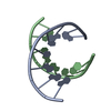
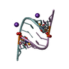
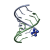
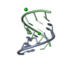
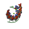
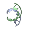
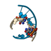
| Title | 3'-DNA-RNA-5' JUNCTION FORMED DURING INITIATION OF MINUS-STRAND SYNTHESIS OF HIV REPLICATION | ||||||
 Components Components |
| ||||||
 Keywords Keywords | DNA-RNA HYBRID / A-DNA/RNA / DOUBLE HELIX | ||||||
| Function / homology | DNA/RNA hybrid / RNA Function and homology information Function and homology information | ||||||
| Method |  X-RAY DIFFRACTION / SYNCHROTRON / Resolution: 1.94 Å X-RAY DIFFRACTION / SYNCHROTRON / Resolution: 1.94 Å | ||||||
 Authors Authors | Mueller, U. / Meier, G. / Mochi-Onori, A. / Cellai, L. / Heumann, H. | ||||||
 Citation Citation | Journal: Biochemistry / Year: 1998 Title: Crystal structure of an eight-base pair duplex containing the 3'-DNA-RNA-5' junction formed during initiation of minus-strand synthesis of HIV replication. Authors: Mueller, U. / Maier, G. / Mochi Onori, A. / Cellai, L. / Heumann, H. / Heinemann, U. | ||||||
| History |
|
- Structure visualization
| Structure viewer | Molecule: MolmilJmol/JSmol |
|---|
- Downloads & links
-Download
| PDBx/mmCIF format | 398d.cif.gz | 29.3 KB | Display | PDBx/mmCIF format |
|---|---|---|---|---|
| PDB format | pdb398d.ent.gz | 18.7 KB | Display | PDB format |
| PDBx/mmJSON format | 398d.json.gz | Tree view | PDBx/mmJSON format | |
| Others |  Other downloads Other downloads |
-Validation report
| Arichive directory | https://data.pdbj.org/pub/pdb/validation_reports/98/398dftp://data.pdbj.org/pub/pdb/validation_reports/98/398d | HTTPS FTP |
|---|
-Related structure data
| Similar structure data |
|---|


-Links
 PDBj
PDBj- Assembly
| Deposited unit | 
| ||||||||||
|---|---|---|---|---|---|---|---|---|---|---|---|
| 1 | 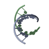
| ||||||||||
| 2 | 
| ||||||||||
| Unit cell |
|
-Components
| #1: RNA chain | Mass: 2581.601 Da / Num. of mol.: 2 / Source method: obtained synthetically #2: DNA/RNA hybrid | Mass: 2451.580 Da / Num. of mol.: 2 / Source method: obtained synthetically #3: Water | ChemComp-HOH / |  Mass: 18.015 Da / Num. of mol.: 84 / Source method: isolated from a natural source / Formula: H2O Mass: 18.015 Da / Num. of mol.: 84 / Source method: isolated from a natural source / Formula: H2O |
|---|
-Experimental details
-Experiment
| Experiment | Method: X-RAY DIFFRACTION / Number of used crystals: 2 |
|---|
- Sample preparation
| Crystal | Density Matthews: 2.43 Å3/Da / Density % sol: 49.46 % | ||||||||||||||||||||||||||||||||||||||||||||||||
|---|---|---|---|---|---|---|---|---|---|---|---|---|---|---|---|---|---|---|---|---|---|---|---|---|---|---|---|---|---|---|---|---|---|---|---|---|---|---|---|---|---|---|---|---|---|---|---|---|---|
| Crystal grow | Method: vapor diffusion, hanging drop / pH: 7 / Details: pH 7.00, VAPOR DIFFUSION, HANGING DROP | ||||||||||||||||||||||||||||||||||||||||||||||||
| Components of the solutions |
| ||||||||||||||||||||||||||||||||||||||||||||||||
| Crystal grow | *PLUS Temperature: 20 ℃ / pH: 7 | ||||||||||||||||||||||||||||||||||||||||||||||||
| Components of the solutions | *PLUS
|
-Data collection
| Diffraction |
| ||||||||||||
|---|---|---|---|---|---|---|---|---|---|---|---|---|---|
| Diffraction source |
| ||||||||||||
| Detector |
| ||||||||||||
| Radiation |
| ||||||||||||
| Radiation wavelength |
| ||||||||||||
| Reflection | Resolution: 1.94→15 Å / Num. obs: 7612 / % possible obs: 99.2 % / Observed criterion σ(I): 0 / Redundancy: 3.7 % / Biso Wilson estimate: 24.44 Å2 / Rmerge(I) obs: 0.075 / Net I/σ(I): 7.4 | ||||||||||||
| Reflection shell | Resolution: 1.94→2.04 Å / Redundancy: 3 % / Rmerge(I) obs: 0.416 / Mean I/σ(I) obs: 2.2 / % possible all: 93.1 | ||||||||||||
| Reflection | *PLUS Highest resolution: 1.94 Å / Lowest resolution: 15 Å / % possible obs: 99.2 % | ||||||||||||
| Reflection shell | *PLUS Lowest resolution: 1.96 Å / % possible obs: 93.1 % |

- Processing
| Software |
| ||||||||||||||||||||||||||||||||||||||||||||||||||||||||||||
|---|---|---|---|---|---|---|---|---|---|---|---|---|---|---|---|---|---|---|---|---|---|---|---|---|---|---|---|---|---|---|---|---|---|---|---|---|---|---|---|---|---|---|---|---|---|---|---|---|---|---|---|---|---|---|---|---|---|---|---|---|---|
| Refinement | Starting model: CANONICAL A-RNA MODEL OF IDENTICAL SIZE AND SEQUENCE Resolution: 1.94→15 Å / Cross valid method: RANDOM IN THIN SHELLS / σ(F): 0
| ||||||||||||||||||||||||||||||||||||||||||||||||||||||||||||
| Displacement parameters | Biso mean: 24.01 Å2 | ||||||||||||||||||||||||||||||||||||||||||||||||||||||||||||
| Refinement step | Cycle: LAST / Resolution: 1.94→15 Å
| ||||||||||||||||||||||||||||||||||||||||||||||||||||||||||||
| Refine LS restraints |
| ||||||||||||||||||||||||||||||||||||||||||||||||||||||||||||
| LS refinement shell | Resolution: 1.94→2.03 Å / Total num. of bins used: 8
| ||||||||||||||||||||||||||||||||||||||||||||||||||||||||||||
| Xplor file | Serial no: 1 / Param file: DNA-RNA-MULTI-ENDO.PARAM / Topol file: DNA-RNA-MULTI-ENDO.TOP | ||||||||||||||||||||||||||||||||||||||||||||||||||||||||||||
| Software | *PLUS Name: X-PLOR / Classification: refinement | ||||||||||||||||||||||||||||||||||||||||||||||||||||||||||||
| Refinement | *PLUS Highest resolution: 1.94 Å / Lowest resolution: 15 Å / σ(F): 0 / % reflection Rfree: 5 % | ||||||||||||||||||||||||||||||||||||||||||||||||||||||||||||
| Solvent computation | *PLUS | ||||||||||||||||||||||||||||||||||||||||||||||||||||||||||||
| Displacement parameters | *PLUS |
