 Movie
Movie Controller

[English] 日本語
 Yorodumi
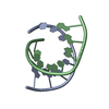
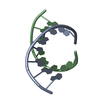
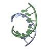
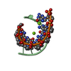
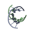
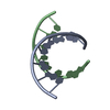
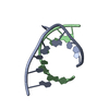
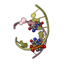
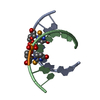
- PDB-370d: STRUCTURAL VARIABILITY OF A-DNA IN CRYSTALS OF THE OCTAMER D(PCPC... -
+ Open data

- Basic information
| Entry | Database: PDB / ID: 370d | ||||||||||||||||||||
|---|---|---|---|---|---|---|---|---|---|---|---|---|---|---|---|---|---|---|---|---|---|
| Title | STRUCTURAL VARIABILITY OF A-DNA IN CRYSTALS OF THE OCTAMER D(PCPCPCPGPCPGPGPG) | ||||||||||||||||||||
 Components Components | DNA (5'-D(P* Keywords KeywordsDNA / A-DNA / DOUBLE HELIX | Function / homology | DNA |  Function and homology information Function and homology informationBiological species | synthetic construct (others) | Method |  X-RAY DIFFRACTION / MOLECULAR REPLACEMENT / Resolution: 1.67 Å X-RAY DIFFRACTION / MOLECULAR REPLACEMENT / Resolution: 1.67 Å  Authors AuthorsFernandez, L.G. / Subirana, J.A. / Verdaguer, N. / Pyshnyi, D. / Campos, L. |  CitationJournal: J.Biomol.Struct.Dyn. / Year: 1997 CitationJournal: J.Biomol.Struct.Dyn. / Year: 1997Title: Structural variability of A-DNA in crystals of the octamer d(pCpCpCpGpCpGpGpG) Authors: Fernandez, L.G. / Subirana, J.A. / Verdaguer, N. / Pyshnyi, D. / Campos, L. / Malinina, L. History |
|
- Structure visualization
| Structure viewer | Molecule: MolmilJmol/JSmol |
|---|
- Downloads & links
-Download
| PDBx/mmCIF format | 370d.cif.gz | 18 KB | Display | PDBx/mmCIF format |
|---|---|---|---|---|
| PDB format | pdb370d.ent.gz | 11.6 KB | Display | PDB format |
| PDBx/mmJSON format | 370d.json.gz | Tree view | PDBx/mmJSON format | |
| Others |  Other downloads Other downloads |
-Validation report
| Arichive directory | https://data.pdbj.org/pub/pdb/validation_reports/70/370dftp://data.pdbj.org/pub/pdb/validation_reports/70/370d | HTTPS FTP |
|---|
-Related structure data


-Links
 PDBj
PDBj

- Assembly
| Deposited unit | 
| ||||||||
|---|---|---|---|---|---|---|---|---|---|
| 1 |
| ||||||||
| Unit cell |
|
-Components
| #1: DNA chain | Mass: 2428.593 Da / Num. of mol.: 2 / Source method: obtained synthetically / Source: (synth.) synthetic construct (others) #2: Chemical | ChemComp-MG / |   Mass: 24.305 Da / Num. of mol.: 1 / Source method: obtained synthetically / Formula: Mg Mass: 24.305 Da / Num. of mol.: 1 / Source method: obtained synthetically / Formula: Mg#3: Water | ChemComp-HOH / |  Mass: 18.015 Da / Num. of mol.: 64 / Source method: isolated from a natural source / Formula: H2O Mass: 18.015 Da / Num. of mol.: 64 / Source method: isolated from a natural source / Formula: H2O |
|---|
-Experimental details
-Experiment
| Experiment | Method: X-RAY DIFFRACTION / Number of used crystals: 1 |
|---|
- Sample preparation
| Crystal | Density Matthews: 2 Å3/Da / Density % sol: 38.57 % | ||||||||||||||||||||||||||||||||||||
|---|---|---|---|---|---|---|---|---|---|---|---|---|---|---|---|---|---|---|---|---|---|---|---|---|---|---|---|---|---|---|---|---|---|---|---|---|---|
| Crystal grow | pH: 6 / Details: pH 6.00 | ||||||||||||||||||||||||||||||||||||
| Components of the solutions |
| ||||||||||||||||||||||||||||||||||||
| Crystal grow | *PLUS Method: vapor diffusion, hanging drop | ||||||||||||||||||||||||||||||||||||
| Components of the solutions | *PLUS
|
-Data collection
| Diffraction | Mean temperature: 293 K |
|---|---|
| Diffraction source | Source: ROTATING ANODE / Type: RIGAKU RU200 |
| Detector | Type: MARRESEARCH / Detector: IMAGE PLATE / Date: Jan 1, 1996 / Details: COLIMATOR 0.4 |
| Radiation | Monochromator: GRAFITO / Monochromatic (M) / Laue (L): M / Scattering type: x-ray |
| Radiation wavelength | Relative weight: 1 |
| Reflection | Resolution: 1.6→20 Å / Num. obs: 4960 / % possible obs: 91.5 % / Redundancy: 9.177 % / Rmerge(I) obs: 0.054 |
| Reflection shell | Resolution: 1.6→1.66 Å / Rmerge(I) obs: 0.1 / % possible all: 80.3 |
| Reflection | *PLUS Highest resolution: 1.6 Å / Lowest resolution: 20 Å / % possible obs: 91.5 % |
| Reflection shell | *PLUS Highest resolution: 1.6 Å / Lowest resolution: 1.66 Å / % possible obs: 80.3 % |
- Processing
| Software |
| ||||||||||||||||||||||||||||||||||||||||||||||||||||||||||||
|---|---|---|---|---|---|---|---|---|---|---|---|---|---|---|---|---|---|---|---|---|---|---|---|---|---|---|---|---|---|---|---|---|---|---|---|---|---|---|---|---|---|---|---|---|---|---|---|---|---|---|---|---|---|---|---|---|---|---|---|---|---|
| Refinement | Method to determine structure: MOLECULAR REPLACEMENT / Resolution: 1.67→8 Å / Cross valid method: R-FREE / σ(F): 2
| ||||||||||||||||||||||||||||||||||||||||||||||||||||||||||||
| Refinement step | Cycle: LAST / Resolution: 1.67→8 Å
| ||||||||||||||||||||||||||||||||||||||||||||||||||||||||||||
| Refine LS restraints |
| ||||||||||||||||||||||||||||||||||||||||||||||||||||||||||||
| LS refinement shell | Resolution: 1.67→1.75 Å / Total num. of bins used: 10
| ||||||||||||||||||||||||||||||||||||||||||||||||||||||||||||
| Xplor file | Serial no: 1 / Param file: PARAM_NDBX_HIGH.DNA / Topol file: TOP_NDBX.DNA | ||||||||||||||||||||||||||||||||||||||||||||||||||||||||||||
| Software | *PLUS Name: X-PLOR / Version: 3.1 / Classification: refinement | ||||||||||||||||||||||||||||||||||||||||||||||||||||||||||||
| Refinement | *PLUS Highest resolution: 1.67 Å / Lowest resolution: 8 Å / σ(F): 2 / % reflection Rfree: 9.51 % / Rfactor obs: 0.181 | ||||||||||||||||||||||||||||||||||||||||||||||||||||||||||||
| Solvent computation | *PLUS | ||||||||||||||||||||||||||||||||||||||||||||||||||||||||||||
| Displacement parameters | *PLUS | ||||||||||||||||||||||||||||||||||||||||||||||||||||||||||||
| Refine LS restraints | *PLUS Type: x_dihedral_angle_deg / Dev ideal: 9.425 | ||||||||||||||||||||||||||||||||||||||||||||||||||||||||||||
| LS refinement shell | *PLUS Rfactor obs: 0.235 |
