 Movie
Movie Controller

[English] 日本語
 Yorodumi
- PDB-5wf5: Agonist bound human A2a adenosine receptor with D52N mutation at ... -
+ Open data

- Basic information
| Entry | Database: PDB / ID: 5wf5 | ||||||
|---|---|---|---|---|---|---|---|
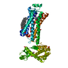
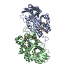
| Title | Agonist bound human A2a adenosine receptor with D52N mutation at 2.60 A resolution | ||||||
 Components Components | Human A2a adenosine receptor T4L chimera | ||||||
 Keywords Keywords | MEMBRANE PROTEIN / human A2a adenosine receptor / D52N / GPCR-T4L chimera / LCP / agonist / UK432097 / Activation microswitch / allosteric Na-site mutant / GPCR signaling | ||||||
| Function / homology |  Function and homology information Function and homology informationregulation of norepinephrine secretion / negative regulation of alpha-beta T cell activation / positive regulation of circadian sleep/wake cycle, sleep / positive regulation of acetylcholine secretion, neurotransmission / Adenosine P1 receptors / G protein-coupled adenosine receptor activity / response to purine-containing compound / G protein-coupled adenosine receptor signaling pathway / NGF-independant TRKA activation / Surfactant metabolism ...regulation of norepinephrine secretion / negative regulation of alpha-beta T cell activation / positive regulation of circadian sleep/wake cycle, sleep / positive regulation of acetylcholine secretion, neurotransmission / Adenosine P1 receptors / G protein-coupled adenosine receptor activity / response to purine-containing compound / G protein-coupled adenosine receptor signaling pathway / NGF-independant TRKA activation / Surfactant metabolism / synaptic transmission, dopaminergic / type 5 metabotropic glutamate receptor binding / negative regulation of vascular permeability / synaptic transmission, cholinergic / intermediate filament / presynaptic active zone / positive regulation of urine volume / response to caffeine / blood circulation / sensory perception / positive regulation of glutamate secretion / eating behavior / inhibitory postsynaptic potential / regulation of calcium ion transport / alpha-actinin binding / asymmetric synapse / axolemma / membrane depolarization / viral release from host cell by cytolysis / cellular defense response / prepulse inhibition / phagocytosis / peptidoglycan catabolic process / positive regulation of synaptic transmission, glutamatergic / neuron projection morphogenesis / astrocyte activation / presynaptic modulation of chemical synaptic transmission / positive regulation of long-term synaptic potentiation / positive regulation of synaptic transmission, GABAergic / central nervous system development / positive regulation of protein secretion / response to amphetamine / regulation of mitochondrial membrane potential / positive regulation of apoptotic signaling pathway / apoptotic signaling pathway / synaptic transmission, glutamatergic / excitatory postsynaptic potential / locomotory behavior / negative regulation of inflammatory response / vasodilation / cell wall macromolecule catabolic process / adenylate cyclase-modulating G protein-coupled receptor signaling pathway / blood coagulation / lysozyme / lysozyme activity / cell-cell signaling / adenylate cyclase-activating G protein-coupled receptor signaling pathway / presynaptic membrane / G alpha (s) signalling events / phospholipase C-activating G protein-coupled receptor signaling pathway / host cell cytoplasm / negative regulation of neuron apoptotic process / calmodulin binding / positive regulation of ERK1 and ERK2 cascade / postsynaptic membrane / defense response to bacterium / response to xenobiotic stimulus / inflammatory response / negative regulation of cell population proliferation / neuronal cell body / apoptotic process / regulation of DNA-templated transcription / lipid binding / dendrite / protein-containing complex binding / glutamatergic synapse / enzyme binding / membrane / identical protein binding / plasma membrane Similarity search - Function | ||||||
| Biological species |  Homo sapiens (human) Homo sapiens (human) Enterobacteria phage T4 (virus) Enterobacteria phage T4 (virus) | ||||||
| Method |  X-RAY DIFFRACTION / SYNCHROTRON / MOLECULAR REPLACEMENT / Resolution: 2.6 Å X-RAY DIFFRACTION / SYNCHROTRON / MOLECULAR REPLACEMENT / Resolution: 2.6 Å | ||||||
 Authors Authors | White, K.L. / Eddy, M.T. / Gao, Z. / Han, G.W. / Hanson, M.A. / Lian, T. / Deary, A. / Patel, N. / Jacobson, K.A. / Katritch, V. / Stevens, R.C. | ||||||
 Citation Citation | Journal: Structure / Year: 2018 Title: Structural Connection between Activation Microswitch and Allosteric Sodium Site in GPCR Signaling. Authors: White, K.L. / Eddy, M.T. / Gao, Z.G. / Han, G.W. / Lian, T. / Deary, A. / Patel, N. / Jacobson, K.A. / Katritch, V. / Stevens, R.C. | ||||||
| History |
|
- Structure visualization
| Structure viewer | Molecule: MolmilJmol/JSmol |
|---|
- Downloads & links
-Download
| PDBx/mmCIF format | 5wf5.cif.gz | 107.1 KB | Display | PDBx/mmCIF format |
|---|---|---|---|---|
| PDB format | pdb5wf5.ent.gz | 77.8 KB | Display | PDB format |
| PDBx/mmJSON format | 5wf5.json.gz | Tree view | PDBx/mmJSON format | |
| Others |  Other downloads Other downloads |
-Validation report
| Arichive directory | https://data.pdbj.org/pub/pdb/validation_reports/wf/5wf5ftp://data.pdbj.org/pub/pdb/validation_reports/wf/5wf5 | HTTPS FTP |
|---|
-Related structure data
| Related structure data |  5wf6C  3emlS S: Starting model for refinement C: citing same article ( |
|---|---|
| Similar structure data |


-Links
 PDBj
PDBj


- Assembly
| Deposited unit | 
| ||||||||
|---|---|---|---|---|---|---|---|---|---|
| 1 |
| ||||||||
| Unit cell |
| ||||||||
| Details | AUTHORS STATE THAT THE BIOLOGICAL UNIT IS UNKNOWN |
-Components
| #1: Protein | Mass: 56555.074 Da / Num. of mol.: 1 / Mutation: R1012G C1054T C1097A I1137R Source method: isolated from a genetically manipulated source Details: D52N mutation Source: (gene. exp.) Homo sapiens (human), (gene. exp.) Enterobacteria phage T4 (virus)Gene: ADORA2A, ADORA2 / Plasmid: pFASTBAC / Production host:   Spodoptera frugiperda (fall armyworm) / Strain (production host): sf9 / References: UniProt: P29274, UniProt: P00720, lysozyme Spodoptera frugiperda (fall armyworm) / Strain (production host): sf9 / References: UniProt: P29274, UniProt: P00720, lysozyme | ||||
|---|---|---|---|---|---|
| #2: Chemical | ChemComp-UKA /   Mass: 777.871 Da / Num. of mol.: 1 / Source method: obtained synthetically / Formula: C40H47N11O6 Mass: 777.871 Da / Num. of mol.: 1 / Source method: obtained synthetically / Formula: C40H47N11O6 | ||||
| #3: Chemical |   Mass: 356.540 Da / Num. of mol.: 2 / Source method: obtained synthetically / Formula: C21H40O4 / Feature type: SUBJECT OF INVESTIGATION Mass: 356.540 Da / Num. of mol.: 2 / Source method: obtained synthetically / Formula: C21H40O4 / Feature type: SUBJECT OF INVESTIGATION#4: Water | ChemComp-HOH / |  Mass: 18.015 Da / Num. of mol.: 3 / Source method: isolated from a natural source / Formula: H2O Mass: 18.015 Da / Num. of mol.: 3 / Source method: isolated from a natural source / Formula: H2OHas protein modification | Y | |
-Experimental details
-Experiment
| Experiment | Method: X-RAY DIFFRACTION / Number of used crystals: 1 |
|---|
- Sample preparation
| Crystal | Density Matthews: 2.88 Å3/Da / Density % sol: 57.22 % |
|---|---|
| Crystal grow | Temperature: 296 K / Method: lipidic cubic phase Details: 100 mM sodium citrate pH 5, 24-27% (v/v) polyethylene glycol (PEG) 400, 30-80 mM MgCl 2 , 5% (v/v) Jeffamine M-600 pH 7 (Hampton) |
-Data collection
| Diffraction | Mean temperature: 100 K |
|---|---|
| Diffraction source | Source: SYNCHROTRON / Site: APS  / Beamline: 23-ID-D / Wavelength: 1.033 Å / Beamline: 23-ID-D / Wavelength: 1.033 Å |
| Detector | Type: DECTRIS PILATUS3 S 6M / Detector: PIXEL / Date: Oct 28, 2016 |
| Radiation | Protocol: SINGLE WAVELENGTH / Monochromatic (M) / Laue (L): M / Scattering type: x-ray |
| Radiation wavelength | Wavelength: 1.033 Å / Relative weight: 1 |
| Reflection | Resolution: 2.6→46.8 Å / Num. obs: 17786 / % possible obs: 92.3 % / Redundancy: 4.4 % / Rmerge(I) obs: 0.18 / Net I/σ(I): 8.1 |
| Reflection shell | Resolution: 2.6→2.69 Å / Redundancy: 3 % / Rmerge(I) obs: 0.88 / Mean I/σ(I) obs: 1.1 / CC1/2: 0.503 / % possible all: 69.4 |
- Processing
| Software |
| |||||||||||||||||||||||||||||||||||||||||||||||||
|---|---|---|---|---|---|---|---|---|---|---|---|---|---|---|---|---|---|---|---|---|---|---|---|---|---|---|---|---|---|---|---|---|---|---|---|---|---|---|---|---|---|---|---|---|---|---|---|---|---|---|
| Refinement | Method to determine structure: MOLECULAR REPLACEMENT Starting model: PDB 3EML Resolution: 2.6→29.909 Å / SU ML: 0.37 / Cross valid method: THROUGHOUT / σ(F): 1.34 / Phase error: 33.94
| |||||||||||||||||||||||||||||||||||||||||||||||||
| Solvent computation | Shrinkage radii: 0.9 Å / VDW probe radii: 1.11 Å | |||||||||||||||||||||||||||||||||||||||||||||||||
| Refinement step | Cycle: LAST / Resolution: 2.6→29.909 Å
| |||||||||||||||||||||||||||||||||||||||||||||||||
| Refine LS restraints |
| |||||||||||||||||||||||||||||||||||||||||||||||||
| LS refinement shell |
|
