[English] 日本語
Yorodumi
- PDB-5w10: Lcd1 GAF domain in complex with cAMP ligand -

+
Open data


ID or keywords:

Loading...

-
Basic information

Entry
Database: PDB / ID: 5w10
TitleLcd1 GAF domain in complex with cAMP ligand
ComponentscGMP-specific phosphodiesterase
KeywordsHYDROLASE / GAF domain / cAMP binding domain / Effector domain / Regulates the DGC activity of the GGDEF domain
Function / homology
Function and homology information


Diguanylate cyclase, GGDEF domain / diguanylate cyclase / GGDEF domain profile. / GGDEF domain / Nucleotide cyclase / GAF domain / Domain present in phytochromes and cGMP-specific phosphodiesterases. / GAF domain / GAF-like domain superfamily / Reverse transcriptase/Diguanylate cyclase domain
Similarity search - Domain/homology
ADENOSINE-3',5'-CYCLIC-MONOPHOSPHATE / cGMP-specific phosphodiesterase
Similarity search - Component
Biological species
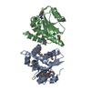
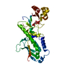
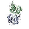
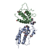
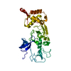
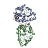
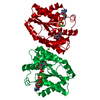
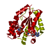
Leptospira interrogans serogroup Icterohaemorrhagiae serovar copenhageni (bacteria)
MethodX-RAY DIFFRACTION / SYNCHROTRON / SAD / Resolution: 2.15 Å
AuthorsGuzzo, C.R. / Maciel, N.K. / Barbosa, A.S. / Farah, C.S.
Funding support Brazil, 4items
OrganizationGrant numberCountry
Sao Paulo Research Foundation (FAPESP)2016/084147 Brazil
Sao Paulo Research Foundation (FAPESP)2013/18664-2 Brazil
Sao Paulo Research Foundation (FAPESP)2013/06650-7 Brazil
Brazilian National Council for Scientific and Technological Development (CNPq)474907/2013-9 Brazil
CitationJournal: J. Mol. Biol. / Year: 2017
Title: Structural and Enzymatic Characterization of a cAMP-Dependent Diguanylate Cyclase from Pathogenic Leptospira Species.
Authors: da Costa Vasconcelos, F.N. / Maciel, N.K. / Favaro, D.C. / de Oliveira, L.C. / Barbosa, A.S. / Salinas, R.K. / de Souza, R.F. / Farah, C.S. / Guzzo, C.R.
History
DepositionJun 1, 2017Deposition site: RCSB / Processing site: RCSB
Revision 1.0Jun 28, 2017Provider: repository / Type: Initial release
Revision 1.1Jul 26, 2017Group: Author supporting evidence / Database references / Category: citation / pdbx_audit_support
Item: _citation.journal_volume / _citation.page_first ..._citation.journal_volume / _citation.page_first / _citation.page_last / _citation.title / _pdbx_audit_support.funding_organization
Revision 1.2Apr 17, 2019Group: Author supporting evidence / Data collection / Category: pdbx_audit_support / Item: _pdbx_audit_support.funding_organization
Revision 1.3Jan 1, 2020Group: Author supporting evidence / Category: pdbx_audit_support / Item: _pdbx_audit_support.funding_organization

-
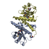
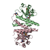
Structure visualization

Structure viewerMolecule:
MolmilJmol/JSmol

Downloads & links

-
Assembly

Deposited unit
A: cGMP-specific phosphodiesterase
B: cGMP-specific phosphodiesterase
C: cGMP-specific phosphodiesterase
D: cGMP-specific phosphodiesterase
hetero molecules


Theoretical massNumber of molelcules
Total (without water)90,2678
Polymers88,9504
Non-polymers1,3174
Water6,233346
1
A: cGMP-specific phosphodiesterase
D: cGMP-specific phosphodiesterase
hetero molecules


Theoretical massNumber of molelcules
Total (without water)45,1334
Polymers44,4752
Non-polymers6582
Water362
TypeNameSymmetry operationNumber
identity operation1_555x,y,z1
Buried area4150 Å2
ΔGint-24 kcal/mol
Surface area16060 Å2
MethodPISA
2
B: cGMP-specific phosphodiesterase
C: cGMP-specific phosphodiesterase
hetero molecules


Theoretical massNumber of molelcules
Total (without water)45,1334
Polymers44,4752
Non-polymers6582
Water362
TypeNameSymmetry operationNumber
identity operation1_555x,y,z1
Buried area4160 Å2
ΔGint-24 kcal/mol
Surface area16090 Å2
MethodPISA
Unit cell
Length a, b, c (Å)119.190, 119.995, 62.454
Angle α, β, γ (deg.)90.00, 90.00, 90.00
Int Tables number18
Space group name H-MP21212
Noncrystallographic symmetry (NCS)NCS domain:
IDEns-IDDetails
11A
21B
12A
22C
13A
23D
14B
24C
15B
25D
16C
26D

NCS domain segments:
Dom-IDComponent-IDEns-IDRefine codeAuth asym-IDAuth seq-ID
1010A4 - 174
2010B4 - 174
1020A3 - 174
2020C3 - 174
1030A3 - 174
2030D3 - 174
1040B4 - 173
2040C4 - 173
1050B4 - 174
2050D4 - 174
1060C1 - 174
2060D1 - 174

NCS ensembles :
ID
1
2
3
4
5
6

-
Components

#1: Protein
cGMP-specific phosphodiesterase


Mass: 22237.525 Da / Num. of mol.: 4
Source method: isolated from a genetically manipulated source
Source: (gene. exp.) Leptospira interrogans serogroup Icterohaemorrhagiae serovar copenhageni (strain Fiocruz L1-130) (bacteria)
Strain: Fiocruz L1-130 / Gene: LIC_13137 / Production host: Escherichia coli (E. coli) / Strain (production host): BL21(DE3)RP / References: UniProt: Q72MQ6
#2: Chemical
ChemComp-CMP / ADENOSINE-3',5'-CYCLIC-MONOPHOSPHATE / CYCLIC AMP / CAMP / Cyclic adenosine monophosphate


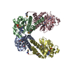
Mass: 329.206 Da / Num. of mol.: 4 / Source method: obtained synthetically / Formula: C10H12N5O6P
#3: Water ChemComp-HOH / water / Water


Mass: 18.015 Da / Num. of mol.: 346 / Source method: isolated from a natural source / Formula: H2O

-
Experimental details

-
Experiment

ExperimentMethod: X-RAY DIFFRACTION / Number of used crystals: 1

-
Sample preparation

CrystalDensity Matthews: 2.54 Å3/Da / Density % sol: 51.62 %
Crystal growTemperature: 291 K / Method: vapor diffusion, sitting drop / Details: 0.2M sodium citrate and 20% PEG 3350

-
Data collection

DiffractionMean temperature: 100 K
Diffraction sourceSource: SYNCHROTRON / Site: LNLS / Beamline: W01B-MX2 / Wavelength: 0.979 Å
DetectorType: MAR CCD 165 mm / Detector: CCD / Date: Apr 16, 2014
RadiationMonochromator: Si(111) double-crystal / Protocol: SINGLE WAVELENGTH / Monochromatic (M) / Laue (L): M / Scattering type: x-ray
Radiation wavelengthWavelength: 0.979 Å / Relative weight: 1
ReflectionResolution: 2.15→40 Å / Num. obs: 93522 / % possible obs: 99.6 % / Redundancy: 3.9 % / Rmerge(I) obs: 0.078 / Net I/σ(I): 17.8
Reflection shellResolution: 2.15→2.23 Å / Redundancy: 2 % / Rmerge(I) obs: 0.56 / Mean I/σ(I) obs: 1.6 / % possible all: 99.2

-
Processing

Software
NameVersionClassification
REFMAC5.7.0032refinement
HKL-2000data reduction
HKL-2000data scaling
CRANK2phasing
RefinementMethod to determine structure: SAD / Resolution: 2.15→29.34 Å / Cor.coef. Fo:Fc: 0.957 / Cor.coef. Fo:Fc free: 0.944 / SU B: 4.944 / SU ML: 0.126 / Cross valid method: THROUGHOUT / ESU R: 0.22 / ESU R Free: 0.177 / Stereochemistry target values: MAXIMUM LIKELIHOOD / Details: HYDROGENS HAVE BEEN ADDED IN THE RIDING POSITIONS
RfactorNum. reflection% reflectionSelection details
Rfree0.22519 2498 5.1 %RANDOM
Rwork0.19463 ---
obs0.19617 46800 99.71 %-
Solvent computationIon probe radii: 0.8 Å / Shrinkage radii: 0.8 Å / VDW probe radii: 1.2 Å / Solvent model: MASK
Displacement parametersBiso mean: 41.894 Å2
Baniso -1Baniso -2Baniso -3
1-0.56 Å20 Å2-0 Å2
2--0.13 Å20 Å2
3----0.69 Å2
Refinement stepCycle: 1 / Resolution: 2.15→29.34 Å
ProteinNucleic acidLigandSolventTotal
Num. atoms5479 0 88 346 5913
Refine LS restraints
Refine-IDTypeDev idealDev ideal targetNumber
X-RAY DIFFRACTIONr_bond_refined_d0.0110.0195814
X-RAY DIFFRACTIONr_bond_other_d0.0070.025640
X-RAY DIFFRACTIONr_angle_refined_deg1.54727881
X-RAY DIFFRACTIONr_angle_other_deg1.4313063
X-RAY DIFFRACTIONr_dihedral_angle_1_deg5.7925722
X-RAY DIFFRACTIONr_dihedral_angle_2_deg35.91724.521261
X-RAY DIFFRACTIONr_dihedral_angle_3_deg15.429151055
X-RAY DIFFRACTIONr_dihedral_angle_4_deg15.5951534
X-RAY DIFFRACTIONr_chiral_restr0.0840.2901
X-RAY DIFFRACTIONr_gen_planes_refined0.0080.026406
X-RAY DIFFRACTIONr_gen_planes_other0.0060.021292
X-RAY DIFFRACTIONr_nbd_refined
X-RAY DIFFRACTIONr_nbd_other
X-RAY DIFFRACTIONr_nbtor_refined
X-RAY DIFFRACTIONr_nbtor_other
X-RAY DIFFRACTIONr_xyhbond_nbd_refined
X-RAY DIFFRACTIONr_xyhbond_nbd_other
X-RAY DIFFRACTIONr_metal_ion_refined
X-RAY DIFFRACTIONr_metal_ion_other
X-RAY DIFFRACTIONr_symmetry_vdw_refined
X-RAY DIFFRACTIONr_symmetry_vdw_other
X-RAY DIFFRACTIONr_symmetry_hbond_refined
X-RAY DIFFRACTIONr_symmetry_hbond_other
X-RAY DIFFRACTIONr_symmetry_metal_ion_refined
X-RAY DIFFRACTIONr_symmetry_metal_ion_other
X-RAY DIFFRACTIONr_mcbond_it3.0573.9032780
X-RAY DIFFRACTIONr_mcbond_other3.0513.9022779
X-RAY DIFFRACTIONr_mcangle_it4.6165.8283475
X-RAY DIFFRACTIONr_mcangle_other4.6155.833476
X-RAY DIFFRACTIONr_scbond_it3.8064.3583034
X-RAY DIFFRACTIONr_scbond_other3.8054.3583035
X-RAY DIFFRACTIONr_scangle_it
X-RAY DIFFRACTIONr_scangle_other5.9396.3364386
X-RAY DIFFRACTIONr_long_range_B_refined8.00631.2156854
X-RAY DIFFRACTIONr_long_range_B_other7.97231.0566721
X-RAY DIFFRACTIONr_rigid_bond_restr
X-RAY DIFFRACTIONr_sphericity_free
X-RAY DIFFRACTIONr_sphericity_bonded
Refine LS restraints NCS

Refine-ID: X-RAY DIFFRACTION / Type: interatomic distance / Weight position: 0.05

Ens-IDDom-IDAuth asym-IDNumberRms dev position (Å)
11A105840.09
12B105840.09
21A95720.16
22C95720.16
31A95200.16
32D95200.16
41B95840.16
42C95840.16
51B94700.16
52D94700.16
61C103300.1
62D103300.1
LS refinement shellResolution: 2.151→2.207 Å / Total num. of bins used: 20
RfactorNum. reflection% reflection
Rfree0.366 185 -
Rwork0.329 3315 -
obs--97.52 %

+
About Yorodumi

-
News

-
Feb 9, 2022. New format data for meta-information of EMDB entries

New format data for meta-information of EMDB entries

  • Version 3 of the EMDB header file is now the official format.
  • The previous official version 1.9 will be removed from the archive.

Related info.:EMDB header

External links:wwPDB to switch to version 3 of the EMDB data model

-
Aug 12, 2020. Covid-19 info

Covid-19 info

URL: https://pdbj.org/emnavi/covid19.php

New page: Covid-19 featured information page in EM Navigator.

Related info.:Covid-19 info / Mar 5, 2020. Novel coronavirus structure data

+
Mar 5, 2020. Novel coronavirus structure data

Novel coronavirus structure data

Related info.:Yorodumi Speices / Aug 12, 2020. Covid-19 info

External links:COVID-19 featured content - PDBj / Molecule of the Month (242):Coronavirus Proteases

+
Jan 31, 2019. EMDB accession codes are about to change! (news from PDBe EMDB page)

EMDB accession codes are about to change! (news from PDBe EMDB page)

  • The allocation of 4 digits for EMDB accession codes will soon come to an end. Whilst these codes will remain in use, new EMDB accession codes will include an additional digit and will expand incrementally as the available range of codes is exhausted. The current 4-digit format prefixed with “EMD-” (i.e. EMD-XXXX) will advance to a 5-digit format (i.e. EMD-XXXXX), and so on. It is currently estimated that the 4-digit codes will be depleted around Spring 2019, at which point the 5-digit format will come into force.
  • The EM Navigator/Yorodumi systems omit the EMD- prefix.

Related info.:Q: What is EMD? / ID/Accession-code notation in Yorodumi/EM Navigator

External links:EMDB Accession Codes are Changing Soon! / Contact to PDBj

+
Jul 12, 2017. Major update of PDB

Major update of PDB

  • wwPDB released updated PDB data conforming to the new PDBx/mmCIF dictionary.
  • This is a major update changing the version number from 4 to 5, and with Remediation, in which all the entries are updated.
  • In this update, many items about electron microscopy experimental information are reorganized (e.g. em_software).
  • Now, EM Navigator and Yorodumi are based on the updated data.

External links:wwPDB Remediation / Enriched Model Files Conforming to OneDep Data Standards Now Available in the PDB FTP Archive

-
Yorodumi

Thousand views of thousand structures

  • Yorodumi is a browser for structure data from EMDB, PDB, SASBDB, etc.
  • This page is also the successor to EM Navigator detail page, and also detail information page/front-end page for Omokage search.
  • The word "yorodu" (or yorozu) is an old Japanese word meaning "ten thousand". "mi" (miru) is to see.

Related info.:EMDB / PDB / SASBDB / Comparison of 3 databanks / Yorodumi Search / Aug 31, 2016. New EM Navigator & Yorodumi / Yorodumi Papers / Jmol/JSmol / Function and homology information / Changes in new EM Navigator and Yorodumi

Read more