 Movie
Movie Controller

[English] 日本語
 Yorodumi
- PDB-4b60: Structure of rFnBPA(189-505) in complex with fibrinogen gamma cha... -
+ Open data

- Basic information
| Entry | Database: PDB / ID: 4b60 | ||||||
|---|---|---|---|---|---|---|---|
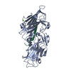
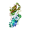
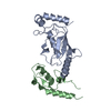
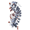
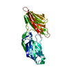
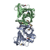
| Title | Structure of rFnBPA(189-505) in complex with fibrinogen gamma chain C- terminal peptide | ||||||
 Components Components |
| ||||||
 Keywords Keywords | CELL ADHESION / FNBPA / FIBRINOGEN / FIBRINOGEN BINDING | ||||||
| Function / homology |  Function and homology information Function and homology informationaggregation of unicellular organisms / fibrinogen complex / platelet alpha granule / Regulation of TLR by endogenous ligand / fibrinogen binding / MyD88 deficiency (TLR2/4) / positive regulation of heterotypic cell-cell adhesion / IRAK4 deficiency (TLR2/4) / MyD88:MAL(TIRAP) cascade initiated on plasma membrane / plasminogen activation ...aggregation of unicellular organisms / fibrinogen complex / platelet alpha granule / Regulation of TLR by endogenous ligand / fibrinogen binding / MyD88 deficiency (TLR2/4) / positive regulation of heterotypic cell-cell adhesion / IRAK4 deficiency (TLR2/4) / MyD88:MAL(TIRAP) cascade initiated on plasma membrane / plasminogen activation / extracellular matrix structural constituent / p130Cas linkage to MAPK signaling for integrins / positive regulation of peptide hormone secretion / GRB2:SOS provides linkage to MAPK signaling for Integrins / fibronectin binding / positive regulation of exocytosis / protein secretion / blood coagulation, fibrin clot formation / protein polymerization / negative regulation of endothelial cell apoptotic process / positive regulation of vasoconstriction / Integrin cell surface interactions / : / negative regulation of extrinsic apoptotic signaling pathway via death domain receptors / fibrinolysis / Integrin signaling / positive regulation of substrate adhesion-dependent cell spreading / cell adhesion molecule binding / platelet alpha granule lumen / cell-matrix adhesion / positive regulation of protein secretion / Post-translational protein phosphorylation / Signaling by high-kinase activity BRAF mutants / MAP2K and MAPK activation / response to calcium ion / platelet aggregation / Regulation of Insulin-like Growth Factor (IGF) transport and uptake by Insulin-like Growth Factor Binding Proteins (IGFBPs) / Signaling by RAF1 mutants / Signaling by moderate kinase activity BRAF mutants / Paradoxical activation of RAF signaling by kinase inactive BRAF / Signaling downstream of RAS mutants / Signaling by BRAF and RAF1 fusions / Platelet degranulation / extracellular matrix / ER-Phagosome pathway / protein-containing complex assembly / blood microparticle / positive regulation of ERK1 and ERK2 cascade / cell adhesion / endoplasmic reticulum lumen / external side of plasma membrane / signaling receptor binding / structural molecule activity / cell surface / : / extracellular exosome / extracellular region / metal ion binding / plasma membrane Similarity search - Function | ||||||
| Biological species |  STAPHYLOCOCCUS AUREUS SUBSP. AUREUS NCTC 8325 (bacteria) STAPHYLOCOCCUS AUREUS SUBSP. AUREUS NCTC 8325 (bacteria) HOMO SAPIENS (human) HOMO SAPIENS (human) | ||||||
| Method |  X-RAY DIFFRACTION / SYNCHROTRON / MOLECULAR REPLACEMENT / Resolution: 1.83 Å X-RAY DIFFRACTION / SYNCHROTRON / MOLECULAR REPLACEMENT / Resolution: 1.83 Å | ||||||
 Authors Authors | Stemberk, V. / Moroz, O. / Atkin, K.E. / Turkenburg, J.P. / Potts, J.R. | ||||||
 Citation Citation | Journal: J.Biol.Chem. / Year: 2014 Title: Evidence for Steric Regulation of Fibrinogen Binding to Staphylococcus Aureus Fibronectin-Binding Protein a (Fnbpa). Authors: Stemberk, V. / Jones, R.P. / Moroz, O. / Atkin, K.E. / Edwards, A.M. / Turkenburg, J.P. / Leech, A.P. / Massey, R.C. / Potts, J.R. | ||||||
| History |
|
- Structure visualization
| Structure viewer | Molecule: MolmilJmol/JSmol |
|---|
- Downloads & links
-Download
| PDBx/mmCIF format | 4b60.cif.gz | 236.9 KB | Display | PDBx/mmCIF format |
|---|---|---|---|---|
| PDB format | pdb4b60.ent.gz | 190.4 KB | Display | PDB format |
| PDBx/mmJSON format | 4b60.json.gz | Tree view | PDBx/mmJSON format | |
| Others |  Other downloads Other downloads |
-Validation report
| Arichive directory | https://data.pdbj.org/pub/pdb/validation_reports/b6/4b60ftp://data.pdbj.org/pub/pdb/validation_reports/b6/4b60 | HTTPS FTP |
|---|
-Related structure data
| Related structure data | 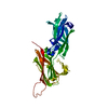 4b5zSC S: Starting model for refinement C: citing same article ( |
|---|---|
| Similar structure data |

-Links
 PDBj
PDBj


- Assembly
| Deposited unit | 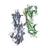
| ||||||||
|---|---|---|---|---|---|---|---|---|---|
| 1 | 
| ||||||||
| 2 | 
| ||||||||
| Unit cell |
|
-Components
| #1: Protein | Mass: 35755.328 Da / Num. of mol.: 2 / Fragment: N2N3, RESIDUES 189-505 Source method: isolated from a genetically manipulated source Source: (gene. exp.) STAPHYLOCOCCUS AUREUS SUBSP. AUREUS NCTC 8325 (bacteria)Production host: #2: Protein/peptide | Mass: 1691.780 Da / Num. of mol.: 2 / Fragment: C-TERMINUS, RESIDUES 421-433 / Source method: obtained synthetically Details: REPRESENTS THE LAST 17 C-TERMINAL RESIDUES OF THE FIBRINOGEN GAMMA CHAIN, ISOFORM GAMMA-A Source: (synth.) HOMO SAPIENS (human) / References: UniProt: P02679#3: Chemical | ChemComp-CA / |   Mass: 40.078 Da / Num. of mol.: 1 / Source method: obtained synthetically / Formula: Ca Mass: 40.078 Da / Num. of mol.: 1 / Source method: obtained synthetically / Formula: Ca#4: Water | ChemComp-HOH / |  Mass: 18.015 Da / Num. of mol.: 406 / Source method: isolated from a natural source / Formula: H2O Mass: 18.015 Da / Num. of mol.: 406 / Source method: isolated from a natural source / Formula: H2OSequence details | GPAM N-TERMINAL RESISDUES FROM VECTOR THE LAST 17 C-TERMINAL RESIDUES OF THE FIBRINOGEN GAMMA ...GPAM N-TERMINAL RESISDUES FROM VECTOR THE LAST 17 C-TERMINAL RESIDUES OF THE FIBRINOGEN | |
|---|
-Experimental details
-Experiment
| Experiment | Method: X-RAY DIFFRACTION / Number of used crystals: 1 |
|---|
- Sample preparation
| Crystal | Density Matthews: 2.23 Å3/Da / Density % sol: 44.93 % / Description: NONE |
|---|---|
| Crystal grow | Details: PEG 2K MME 25%, 0.2 M CA AC, ISOPROPANOL 10% |
-Data collection
| Diffraction | Mean temperature: 100 K |
|---|---|
| Diffraction source | Source: SYNCHROTRON / Site: Diamond  / Beamline: I04-1 / Wavelength: 0.91731 / Beamline: I04-1 / Wavelength: 0.91731 |
| Detector | Type: ADSC CCD / Detector: CCD / Date: Oct 22, 2010 |
| Radiation | Protocol: SINGLE WAVELENGTH / Monochromatic (M) / Laue (L): M / Scattering type: x-ray |
| Radiation wavelength | Wavelength: 0.91731 Å / Relative weight: 1 |
| Reflection | Resolution: 1.8→58.4 Å / Num. obs: 51268 / % possible obs: 93.6 % / Redundancy: 2.2 % / Rmerge(I) obs: 0.07 / Net I/σ(I): 9.1 |
| Reflection shell | Resolution: 1.83→1.87 Å / Redundancy: 2.2 % / Rmerge(I) obs: 0.36 / Mean I/σ(I) obs: 2 / % possible all: 67.4 |
- Processing
| Software |
| ||||||||||||||||||||||||||||||||||||||||||||||||||||||||||||||||||||||||||||||||||||||||||||||||||||||||||||||||||||||||||||||||||||||||||||||||||||||||||||||||||||||||||||||||||||||
|---|---|---|---|---|---|---|---|---|---|---|---|---|---|---|---|---|---|---|---|---|---|---|---|---|---|---|---|---|---|---|---|---|---|---|---|---|---|---|---|---|---|---|---|---|---|---|---|---|---|---|---|---|---|---|---|---|---|---|---|---|---|---|---|---|---|---|---|---|---|---|---|---|---|---|---|---|---|---|---|---|---|---|---|---|---|---|---|---|---|---|---|---|---|---|---|---|---|---|---|---|---|---|---|---|---|---|---|---|---|---|---|---|---|---|---|---|---|---|---|---|---|---|---|---|---|---|---|---|---|---|---|---|---|---|---|---|---|---|---|---|---|---|---|---|---|---|---|---|---|---|---|---|---|---|---|---|---|---|---|---|---|---|---|---|---|---|---|---|---|---|---|---|---|---|---|---|---|---|---|---|---|---|---|
| Refinement | Method to determine structure: MOLECULAR REPLACEMENT Starting model: PDB ENTRY 4B5Z Resolution: 1.83→58.41 Å / Cor.coef. Fo:Fc: 0.947 / Cor.coef. Fo:Fc free: 0.923 / SU B: 6.414 / SU ML: 0.11 / Cross valid method: THROUGHOUT / ESU R: 0.163 / ESU R Free: 0.153 / Stereochemistry target values: MAXIMUM LIKELIHOOD Details: HYDROGENS HAVE BEEN ADDED IN THE RIDING POSITIONS. RESIDUES 189-194, 479-489 AND 504-505 FROM CHAINS A AND B ARE ABSENT AND RESIDUES 1-3 FORM THE C CHAIN AND 1-5 FROM THE D CHAIN ARE ABSENT
| ||||||||||||||||||||||||||||||||||||||||||||||||||||||||||||||||||||||||||||||||||||||||||||||||||||||||||||||||||||||||||||||||||||||||||||||||||||||||||||||||||||||||||||||||||||||
| Solvent computation | Ion probe radii: 0.8 Å / Shrinkage radii: 0.8 Å / VDW probe radii: 1.2 Å / Solvent model: MASK | ||||||||||||||||||||||||||||||||||||||||||||||||||||||||||||||||||||||||||||||||||||||||||||||||||||||||||||||||||||||||||||||||||||||||||||||||||||||||||||||||||||||||||||||||||||||
| Displacement parameters | Biso mean: 24.17 Å2
| ||||||||||||||||||||||||||||||||||||||||||||||||||||||||||||||||||||||||||||||||||||||||||||||||||||||||||||||||||||||||||||||||||||||||||||||||||||||||||||||||||||||||||||||||||||||
| Refinement step | Cycle: LAST / Resolution: 1.83→58.41 Å
| ||||||||||||||||||||||||||||||||||||||||||||||||||||||||||||||||||||||||||||||||||||||||||||||||||||||||||||||||||||||||||||||||||||||||||||||||||||||||||||||||||||||||||||||||||||||
| Refine LS restraints |
|