 Movie
Movie Controller


+ Open data

- Basic information
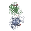
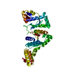
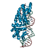
| Entry | Database: PDB / ID: 1eqn | ||||||
|---|---|---|---|---|---|---|---|
| Title | E.COLI PRIMASE CATALYTIC CORE | ||||||
 Components Components | DNA PRIMASE | ||||||
 Keywords Keywords | TRANSFERASE / Toprim domain / Rossmann fold | ||||||
| Function / homology |  Function and homology information Function and homology informationDnaB-DnaG complex / DNA primase DnaG / primosome complex / DNA replication, synthesis of primer / replisome / replication fork processing / DNA-directed RNA polymerase complex / DNA-directed RNA polymerase activity / DNA replication / DNA binding ...DnaB-DnaG complex / DNA primase DnaG / primosome complex / DNA replication, synthesis of primer / replisome / replication fork processing / DNA-directed RNA polymerase complex / DNA-directed RNA polymerase activity / DNA replication / DNA binding / zinc ion binding / cytoplasm Similarity search - Function | ||||||
| Biological species |  | ||||||
| Method |  X-RAY DIFFRACTION / SYNCHROTRON / Resolution: 2.9 Å X-RAY DIFFRACTION / SYNCHROTRON / Resolution: 2.9 Å | ||||||
 Authors Authors | Podobnik, M. / McInerney, P. / O'Donnell, M. / Kuriyan, J. | ||||||
 Citation Citation | Journal: J.Mol.Biol. / Year: 2000 Title: A TOPRIM domain in the crystal structure of the catalytic core of Escherichia coli primase confirms a structural link to DNA topoisomerases. Authors: Podobnik, M. / McInerney, P. / O'Donnell, M. / Kuriyan, J. | ||||||
| History |
|
- Structure visualization
| Structure viewer | Molecule: MolmilJmol/JSmol |
|---|
- Downloads & links
-Download
| PDBx/mmCIF format | 1eqn.cif.gz | 306.8 KB | Display | PDBx/mmCIF format |
|---|---|---|---|---|
| PDB format | pdb1eqn.ent.gz | 251.9 KB | Display | PDB format |
| PDBx/mmJSON format | 1eqn.json.gz | Tree view | PDBx/mmJSON format | |
| Others |  Other downloads Other downloads |
-Validation report
| Arichive directory | https://data.pdbj.org/pub/pdb/validation_reports/eq/1eqnftp://data.pdbj.org/pub/pdb/validation_reports/eq/1eqn | HTTPS FTP |
|---|
-Related structure data
| Similar structure data |
|---|


-Links
 PDBj
PDBj

- Assembly
| Deposited unit | 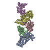
| ||||||||
|---|---|---|---|---|---|---|---|---|---|
| 1 | 
| ||||||||
| 2 | 
| ||||||||
| 3 | 
| ||||||||
| 4 | 
| ||||||||
| 5 | 
| ||||||||
| Unit cell |
| ||||||||
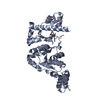
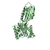
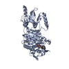
| Details | The biological assembly is a monomer constructed from either chain A, B, C, D or E. |
-Components
| #1: Protein | Mass: 36620.941 Da / Num. of mol.: 5 / Fragment: CATALYTIC CORE Source method: isolated from a genetically manipulated source Source: (gene. exp.) References: UniProt: P0ABS5, Transferases; Transferring phosphorus-containing groups; Nucleotidyltransferases Has protein modification | Y | |
|---|
-Experimental details
-Experiment
| Experiment | Method: X-RAY DIFFRACTION / Number of used crystals: 1 |
|---|
- Sample preparation
| Crystal | Density Matthews: 2.42 Å3/Da / Density % sol: 49.21 % Description: For the refinement the data from lambda 1 (0.98636) were taken. | |||||||||||||||||||||||||
|---|---|---|---|---|---|---|---|---|---|---|---|---|---|---|---|---|---|---|---|---|---|---|---|---|---|---|
| Crystal grow | Temperature: 293 K / Method: vapor diffusion, hanging drop / pH: 8.3 Details: PEG 4000, DTT, Tris/HCl, sodium acetate, pH 8.3, VAPOR DIFFUSION, HANGING DROP, temperature 293K | |||||||||||||||||||||||||
| Crystal | *PLUS Density % sol: 50 % | |||||||||||||||||||||||||
| Crystal grow | *PLUS Temperature: 20 ℃Details: drop consists of equal volume of protein and reservoir solutions PH range low: 8.5 / PH range high: 7.9 | |||||||||||||||||||||||||
| Components of the solutions | *PLUS
|
-Data collection
| Diffraction | Mean temperature: 100 K | |||||||||||||||
|---|---|---|---|---|---|---|---|---|---|---|---|---|---|---|---|---|
| Diffraction source | Source: SYNCHROTRON / Site: NSLS  / Beamline: X4A / Wavelength: 0.98636, 0.97939, 0.97900, 0.96526 / Beamline: X4A / Wavelength: 0.98636, 0.97939, 0.97900, 0.96526 | |||||||||||||||
| Detector | Type: ADSC QUANTUM 4 / Detector: CCD / Date: Jun 1, 1999 | |||||||||||||||
| Radiation | Protocol: MAD / Monochromatic (M) / Laue (L): M / Scattering type: x-ray | |||||||||||||||
| Radiation wavelength |
| |||||||||||||||
| Reflection | Resolution: 2.9→20 Å / Num. all: 273234 / Num. obs: 272858 / % possible obs: 97.6 % / Observed criterion σ(F): -1.5 / Observed criterion σ(I): -3 / Redundancy: 6.9 % / Biso Wilson estimate: 73.637 Å2 / Rmerge(I) obs: 0.058 / Net I/σ(I): 31.3 | |||||||||||||||
| Reflection shell | Resolution: 2.9→3 Å / Redundancy: 3.6 % / Rmerge(I) obs: 0.207 / Num. unique all: 13126 / % possible all: 90.8 | |||||||||||||||
| Reflection | *PLUS Num. obs: 40874 / Num. measured all: 273234 | |||||||||||||||
| Reflection shell | *PLUS % possible obs: 90.8 % |
- Processing
| Software |
| |||||||||||||||||||||||||
|---|---|---|---|---|---|---|---|---|---|---|---|---|---|---|---|---|---|---|---|---|---|---|---|---|---|---|
| Refinement | Resolution: 2.9→500 Å / σ(F): 2 Stereochemistry target values: Engh & Huber, Hendrickson W.A. and Konnert J.H. Details: used standard crystallographic residual
| |||||||||||||||||||||||||
| Refinement step | Cycle: LAST / Resolution: 2.9→500 Å
| |||||||||||||||||||||||||
| Refine LS restraints |
|
