 Movie
Movie Controller

[English] 日本語
 Yorodumi
- PDB-5vtl: Structure of metacyclic invariant surface protein , Tb427.07.360,... -
+ Open data

- Basic information
| Entry | Database: PDB / ID: 5vtl | ||||||
|---|---|---|---|---|---|---|---|
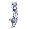
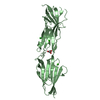
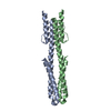
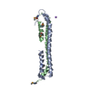
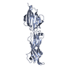
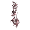
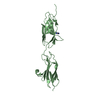
| Title | Structure of metacyclic invariant surface protein , Tb427.07.360, from Trypanosoma brucei. | ||||||
 Components Components | Tb427.07.360- putative uncharacterized metacyclic invariant surface protein from Trypanosoma brucei | ||||||
 Keywords Keywords | UNKNOWN FUNCTION / Extended helical architecture / invariant surface protein | ||||||
| Function / homology | Trypanosoma glutamic acid/alanine-rich protein / Glutamic acid/alanine-rich protein of Trypanosoma / Tb427.07.360- putative uncharacterized metacyclic invariant surface protein from Trypanosoma brucei Function and homology information Function and homology information | ||||||
| Biological species |  | ||||||
| Method |  X-RAY DIFFRACTION / SYNCHROTRON / MOLECULAR REPLACEMENT / Resolution: 1.8231 Å X-RAY DIFFRACTION / SYNCHROTRON / MOLECULAR REPLACEMENT / Resolution: 1.8231 Å | ||||||
 Authors Authors | Ramswamy, R. / Workman, S.D. / Boulanger, M.J. | ||||||
| Funding support |  Canada, 1items Canada, 1items
| ||||||
 Citation Citation | Journal: to be published Title: Structure of Tb427.07.360, an insect stage antigen from Trypanosoma brucei. Authors: Ramswamy, R. / Workman, S.D. / Boulanger, M.J. | ||||||
| History |
|
- Structure visualization
| Structure viewer | Molecule: MolmilJmol/JSmol |
|---|
- Downloads & links
-Download
| PDBx/mmCIF format | 5vtl.cif.gz | 90.7 KB | Display | PDBx/mmCIF format |
|---|---|---|---|---|
| PDB format | pdb5vtl.ent.gz | 67.8 KB | Display | PDB format |
| PDBx/mmJSON format | 5vtl.json.gz | Tree view | PDBx/mmJSON format | |
| Others |  Other downloads Other downloads |
-Validation report
| Arichive directory | https://data.pdbj.org/pub/pdb/validation_reports/vt/5vtlftp://data.pdbj.org/pub/pdb/validation_reports/vt/5vtl | HTTPS FTP |
|---|
-Related structure data
| Related structure data | 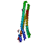 2y44S S: Starting model for refinement |
|---|---|
| Similar structure data |


-Links
 PDBj
PDBj- Assembly
| Deposited unit | 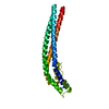
| ||||||||
|---|---|---|---|---|---|---|---|---|---|
| 1 |
| ||||||||
| Unit cell |
|
-Components
| #1: Protein | Mass: 22087.674 Da / Num. of mol.: 1 Source method: isolated from a genetically manipulated source Source: (gene. exp.)  |
|---|---|
| #2: Water | ChemComp-HOH /  Mass: 18.015 Da / Num. of mol.: 273 / Source method: isolated from a natural source / Formula: H2O Mass: 18.015 Da / Num. of mol.: 273 / Source method: isolated from a natural source / Formula: H2O |
| Has protein modification | Y |
-Experimental details
-Experiment
| Experiment | Method: X-RAY DIFFRACTION / Number of used crystals: 1 |
|---|
- Sample preparation
| Crystal | Density Matthews: 2.41 Å3/Da / Density % sol: 49.07 % |
|---|---|
| Crystal grow | Temperature: 291 K / Method: vapor diffusion, sitting drop / Details: 25% PEG 1500 |
-Data collection
| Diffraction | Mean temperature: 100 K |
|---|---|
| Diffraction source | Source: SYNCHROTRON / Site: SSRL  / Beamline: BL7-1 / Wavelength: 0.9795 Å / Beamline: BL7-1 / Wavelength: 0.9795 Å |
| Detector | Type: ADSC QUANTUM 315r / Detector: CCD / Date: Nov 1, 2014 / Details: Rh coated focusing mirror |
| Radiation | Monochromator: Side scattering I-beam bent single crystal; asymmetric cut 4.9650 deg Protocol: SINGLE WAVELENGTH / Monochromatic (M) / Laue (L): M / Scattering type: x-ray |
| Radiation wavelength | Wavelength: 0.9795 Å / Relative weight: 1 |
| Reflection | Resolution: 1.82→44.72 Å / Num. obs: 136043 / % possible obs: 98.2 % / Redundancy: 6.9 % / Biso Wilson estimate: 12.25 Å2 / Net I/σ(I): 20.8 |
| Reflection shell | Resolution: 1.82→1.92 Å / Redundancy: 6.9 % / Num. unique obs: 19605 / % possible all: 98.2 |
- Processing
| Software |
| ||||||||||||||||||||||||||||||||||||||||||||||||||||||||
|---|---|---|---|---|---|---|---|---|---|---|---|---|---|---|---|---|---|---|---|---|---|---|---|---|---|---|---|---|---|---|---|---|---|---|---|---|---|---|---|---|---|---|---|---|---|---|---|---|---|---|---|---|---|---|---|---|---|
| Refinement | Method to determine structure: MOLECULAR REPLACEMENT Starting model: 2Y44 Resolution: 1.8231→44.72 Å / SU ML: 0.18 / Cross valid method: FREE R-VALUE / σ(F): 1.36 / Phase error: 18.04
| ||||||||||||||||||||||||||||||||||||||||||||||||||||||||
| Solvent computation | Shrinkage radii: 0.9 Å / VDW probe radii: 1.11 Å | ||||||||||||||||||||||||||||||||||||||||||||||||||||||||
| Displacement parameters | Biso max: 86.03 Å2 / Biso mean: 12.3 Å2 / Biso min: 2.2 Å2 | ||||||||||||||||||||||||||||||||||||||||||||||||||||||||
| Refinement step | Cycle: final / Resolution: 1.8231→44.72 Å
| ||||||||||||||||||||||||||||||||||||||||||||||||||||||||
| Refine LS restraints |
| ||||||||||||||||||||||||||||||||||||||||||||||||||||||||
| LS refinement shell | Refine-ID: X-RAY DIFFRACTION / Rfactor Rfree error: 0 / Total num. of bins used: 7
|
