 Movie
Movie Controller

[English] 日本語
 Yorodumi
- PDB-5va6: CRYSTAL STRUCTURE OF ATXR5 IN COMPLEX WITH HISTONE H3.1 MONO-METH... -
+ Open data

- Basic information
| Entry | Database: PDB / ID: 5va6 | ||||||
|---|---|---|---|---|---|---|---|
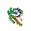
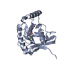
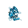
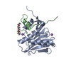
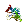
| Title | CRYSTAL STRUCTURE OF ATXR5 IN COMPLEX WITH HISTONE H3.1 MONO-METHYLATED ON R26 | ||||||
 Components Components |
| ||||||
 Keywords Keywords | TRANSFERASE/DNA BINDING PROTEIN / nucleosome / TRANSFERASE-DNA BINDING PROTEIN complex | ||||||
| Function / homology |  Function and homology information Function and homology information[histone H3]-lysine27 N-methyltransferase / histone H3K27 monomethyltransferase activity / histone acetyltransferase activity / regulation of DNA replication / Chromatin modifying enzymes / epigenetic regulation of gene expression / telomere organization / Interleukin-7 signaling / RNA Polymerase I Promoter Opening / Assembly of the ORC complex at the origin of replication ...[histone H3]-lysine27 N-methyltransferase / histone H3K27 monomethyltransferase activity / histone acetyltransferase activity / regulation of DNA replication / Chromatin modifying enzymes / epigenetic regulation of gene expression / telomere organization / Interleukin-7 signaling / RNA Polymerase I Promoter Opening / Assembly of the ORC complex at the origin of replication / Regulation of endogenous retroelements by the Human Silencing Hub (HUSH) complex / DNA methylation / Condensation of Prophase Chromosomes / Chromatin modifications during the maternal to zygotic transition (MZT) / SIRT1 negatively regulates rRNA expression / HCMV Late Events / ERCC6 (CSB) and EHMT2 (G9a) positively regulate rRNA expression / PRC2 methylates histones and DNA / Regulation of endogenous retroelements by KRAB-ZFP proteins / chloroplast / Defective pyroptosis / transcription coregulator activity / HDACs deacetylate histones / Regulation of endogenous retroelements by Piwi-interacting RNAs (piRNAs) / RNA Polymerase I Promoter Escape / Transcriptional regulation by small RNAs / HDMs demethylate histones / Formation of the beta-catenin:TCF transactivating complex / Activated PKN1 stimulates transcription of AR (androgen receptor) regulated genes KLK2 and KLK3 / RUNX1 regulates genes involved in megakaryocyte differentiation and platelet function / NoRC negatively regulates rRNA expression / Negative Regulation of CDH1 Gene Transcription / PKMTs methylate histone lysines / B-WICH complex positively regulates rRNA expression / Meiotic recombination / Pre-NOTCH Transcription and Translation / Activation of anterior HOX genes in hindbrain development during early embryogenesis / Transcriptional regulation of granulopoiesis / RMTs methylate histone arginines / HCMV Early Events / structural constituent of chromatin / nucleosome / nucleosome assembly / HATs acetylate histones / Factors involved in megakaryocyte development and platelet production / MLL4 and MLL3 complexes regulate expression of PPARG target genes in adipogenesis and hepatic steatosis / chromatin organization / RUNX1 regulates transcription of genes involved in differentiation of HSCs / Senescence-Associated Secretory Phenotype (SASP) / methylation / Oxidative Stress Induced Senescence / Estrogen-dependent gene expression / regulation of cell cycle / cadherin binding / Amyloid fiber formation / protein heterodimerization activity / chromatin binding / regulation of transcription by RNA polymerase II / chromatin / protein-containing complex / DNA binding / extracellular exosome / extracellular region / zinc ion binding / nucleoplasm / membrane / nucleus Similarity search - Function | ||||||
| Biological species |  Ricinus communis (castor bean) Ricinus communis (castor bean) Homo sapiens (human) Homo sapiens (human) | ||||||
| Method |  X-RAY DIFFRACTION / SYNCHROTRON / MOLECULAR REPLACEMENT / Resolution: 2.4 Å X-RAY DIFFRACTION / SYNCHROTRON / MOLECULAR REPLACEMENT / Resolution: 2.4 Å | ||||||
 Authors Authors | Bergamin, E. / Sarvan, S. / Malette, J. / Eram, M. / Yeung, S. / Mongeon, V. / Joshi, M. / Brunzelle, J.S. / Michaels, S.D. / Blais, A. ...Bergamin, E. / Sarvan, S. / Malette, J. / Eram, M. / Yeung, S. / Mongeon, V. / Joshi, M. / Brunzelle, J.S. / Michaels, S.D. / Blais, A. / Vedadi, M. / Couture, J.-F. | ||||||
 Citation Citation | Journal: Nucleic Acids Res. / Year: 2017 Title: Molecular basis for the methylation specificity of ATXR5 for histone H3. Authors: Bergamin, E. / Sarvan, S. / Malette, J. / Eram, M.S. / Yeung, S. / Mongeon, V. / Joshi, M. / Brunzelle, J.S. / Michaels, S.D. / Blais, A. / Vedadi, M. / Couture, J.F. | ||||||
| History |
|
- Structure visualization
| Structure viewer | Molecule: MolmilJmol/JSmol |
|---|
- Downloads & links
-Download
| PDBx/mmCIF format | 5va6.cif.gz | 189.2 KB | Display | PDBx/mmCIF format |
|---|---|---|---|---|
| PDB format | pdb5va6.ent.gz | 148.2 KB | Display | PDB format |
| PDBx/mmJSON format | 5va6.json.gz | Tree view | PDBx/mmJSON format | |
| Others |  Other downloads Other downloads |
-Validation report
| Arichive directory | https://data.pdbj.org/pub/pdb/validation_reports/va/5va6ftp://data.pdbj.org/pub/pdb/validation_reports/va/5va6 | HTTPS FTP |
|---|
-Related structure data
| Related structure data |  5vabC  5vacC  5vahC  5vbcC  4o30S S: Starting model for refinement C: citing same article ( |
|---|---|
| Similar structure data |


-Links
 PDBj
PDBj


- Assembly
| Deposited unit | 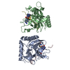
| ||||||||
|---|---|---|---|---|---|---|---|---|---|
| 1 | 
| ||||||||
| 2 | 
| ||||||||
| Unit cell |
|
-Components
| #1: Protein | Mass: 26218.723 Da / Num. of mol.: 2 / Fragment: residues 146-374 Source method: isolated from a genetically manipulated source Source: (gene. exp.) Ricinus communis (castor bean) / Gene: ATXR5, RCOM_1460410 / Production host:  References: UniProt: B9RU15, histone-lysine N-methyltransferase #2: Protein/peptide | Mass: 1772.078 Da / Num. of mol.: 2 / Fragment: residues 20-37 / Source method: obtained synthetically / Source: (synth.) Homo sapiens (human) / References: UniProt: P68431#3: Chemical |   Type: L-peptide linking / Mass: 384.411 Da / Num. of mol.: 2 / Source method: obtained synthetically / Formula: C14H20N6O5S Type: L-peptide linking / Mass: 384.411 Da / Num. of mol.: 2 / Source method: obtained synthetically / Formula: C14H20N6O5S#4: Water | ChemComp-HOH / |  Mass: 18.015 Da / Num. of mol.: 159 / Source method: isolated from a natural source / Formula: H2O Mass: 18.015 Da / Num. of mol.: 159 / Source method: isolated from a natural source / Formula: H2O |
|---|
-Experimental details
-Experiment
| Experiment | Method: X-RAY DIFFRACTION / Number of used crystals: 1 |
|---|
- Sample preparation
| Crystal | Density Matthews: 2.48 Å3/Da / Density % sol: 50.42 % |
|---|---|
| Crystal grow | Temperature: 277 K / Method: vapor diffusion, hanging drop Details: 50% polypropylene glycol 400, 5% DMSO, 0.1 M HEPES-NaOH (pH 6.0) |
-Data collection
| Diffraction | Mean temperature: 100 K |
|---|---|
| Diffraction source | Source: SYNCHROTRON / Site: APS  / Beamline: 17-ID / Wavelength: 1 Å / Beamline: 17-ID / Wavelength: 1 Å |
| Detector | Type: MARMOSAIC 300 mm CCD / Detector: CCD / Date: Mar 1, 2014 |
| Radiation | Protocol: SINGLE WAVELENGTH / Monochromatic (M) / Laue (L): M / Scattering type: x-ray |
| Radiation wavelength | Wavelength: 1 Å / Relative weight: 1 |
| Reflection | Resolution: 2.4→17 Å / Num. obs: 17859 / % possible obs: 96.1 % / Redundancy: 3.9 % / Biso Wilson estimate: 55.8 Å2 / Rsym value: 0.061 / Net I/σ(I): 7.8 |
- Processing
| Software |
| ||||||||||||||||||||||||||||||||||||||||||||||||||||||||||||||||||||||||||||||||||||||||||||||||||||||||||||||||||
|---|---|---|---|---|---|---|---|---|---|---|---|---|---|---|---|---|---|---|---|---|---|---|---|---|---|---|---|---|---|---|---|---|---|---|---|---|---|---|---|---|---|---|---|---|---|---|---|---|---|---|---|---|---|---|---|---|---|---|---|---|---|---|---|---|---|---|---|---|---|---|---|---|---|---|---|---|---|---|---|---|---|---|---|---|---|---|---|---|---|---|---|---|---|---|---|---|---|---|---|---|---|---|---|---|---|---|---|---|---|---|---|---|---|---|---|
| Refinement | Method to determine structure: MOLECULAR REPLACEMENT Starting model: 4O30 Resolution: 2.4→16.57 Å / Cor.coef. Fo:Fc: 0.9057 / Cor.coef. Fo:Fc free: 0.8661 / SU R Cruickshank DPI: 0.678 / Cross valid method: THROUGHOUT / σ(F): 0 / SU R Blow DPI: 0.685 / SU Rfree Blow DPI: 0.331 / SU Rfree Cruickshank DPI: 0.335
| ||||||||||||||||||||||||||||||||||||||||||||||||||||||||||||||||||||||||||||||||||||||||||||||||||||||||||||||||||
| Displacement parameters | Biso mean: 35.32 Å2
| ||||||||||||||||||||||||||||||||||||||||||||||||||||||||||||||||||||||||||||||||||||||||||||||||||||||||||||||||||
| Refine analyze | Luzzati coordinate error obs: 0.422 Å | ||||||||||||||||||||||||||||||||||||||||||||||||||||||||||||||||||||||||||||||||||||||||||||||||||||||||||||||||||
| Refinement step | Cycle: 1 / Resolution: 2.4→16.57 Å
| ||||||||||||||||||||||||||||||||||||||||||||||||||||||||||||||||||||||||||||||||||||||||||||||||||||||||||||||||||
| Refine LS restraints |
| ||||||||||||||||||||||||||||||||||||||||||||||||||||||||||||||||||||||||||||||||||||||||||||||||||||||||||||||||||
| LS refinement shell | Resolution: 2.4→2.6 Å / Total num. of bins used: 9
| ||||||||||||||||||||||||||||||||||||||||||||||||||||||||||||||||||||||||||||||||||||||||||||||||||||||||||||||||||
| Refinement TLS params. | Method: refined / Refine-ID: X-RAY DIFFRACTION
| ||||||||||||||||||||||||||||||||||||||||||||||||||||||||||||||||||||||||||||||||||||||||||||||||||||||||||||||||||
| Refinement TLS group |
|
