 Movie
Movie Controller

[English] 日本語
 Yorodumi
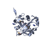
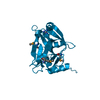
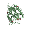
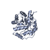
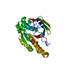
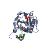
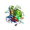
- PDB-4izj: Crystal structure of yellowtail ascites virus VP4 protease with a... -
+ Open data

- Basic information
| Entry | Database: PDB / ID: 4izj | |||||||||
|---|---|---|---|---|---|---|---|---|---|---|
| Title | Crystal structure of yellowtail ascites virus VP4 protease with a wild-type active site reveals acyl-enzyme complexes and product complexes. | |||||||||
 Components Components | (Yellowtail Ascites Virus (YAV) VP4 ...) x 3 | |||||||||
 Keywords Keywords | HYDROLASE / VIRAL PROTEASE / BIRNAVIRUS / SERINE-LYSINE DYAD MECHANISM / ALPHA-BETA PROTEIN FOLD / LYSINE GENERAL BASE / ACYL-ENZYME / PRODUCT COMPLEX | |||||||||
| Function / homology |  Function and homology information Function and homology informationHydrolases; Acting on peptide bonds (peptidases); Serine endopeptidases / serine-type peptidase activity / viral capsid / host cell cytoplasm / structural molecule activity / proteolysis / metal ion binding Similarity search - Function | |||||||||
| Biological species |  Yellowtail ascites virus Yellowtail ascites virus | |||||||||
| Method |  X-RAY DIFFRACTION / MOLECULAR REPLACEMENT / Resolution: 2.5 Å X-RAY DIFFRACTION / MOLECULAR REPLACEMENT / Resolution: 2.5 Å | |||||||||
 Authors Authors | Paetzel, M. / Chung, I.Y.W. | |||||||||
 Citation Citation | Journal: J.Biol.Chem. / Year: 2013 Title: Crystal Structures of Yellowtail Ascites Virus VP4 Protease: TRAPPING AN INTERNAL CLEAVAGE SITE TRANS ACYL-ENZYME COMPLEX IN A NATIVE SER/LYS DYAD ACTIVE SITE. Authors: Chung, I.Y. / Paetzel, M. | |||||||||
| History |
|
- Structure visualization
| Structure viewer | Molecule: MolmilJmol/JSmol |
|---|
- Downloads & links
-Download
| PDBx/mmCIF format | 4izj.cif.gz | 203.9 KB | Display | PDBx/mmCIF format |
|---|---|---|---|---|
| PDB format | pdb4izj.ent.gz | 162.6 KB | Display | PDB format |
| PDBx/mmJSON format | 4izj.json.gz | Tree view | PDBx/mmJSON format | |
| Others |  Other downloads Other downloads |
-Validation report
| Arichive directory | https://data.pdbj.org/pub/pdb/validation_reports/iz/4izjftp://data.pdbj.org/pub/pdb/validation_reports/iz/4izj | HTTPS FTP |
|---|
-Related structure data
| Related structure data | 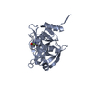 4izkC 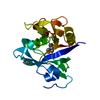 2pnmS C: citing same article ( S: Starting model for refinement |
|---|---|
| Similar structure data |


-Links
 PDBj
PDBj


- Assembly
| Deposited unit | 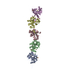
| ||||||||
|---|---|---|---|---|---|---|---|---|---|
| 1 | 
| ||||||||
| 2 | 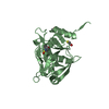
| ||||||||
| 3 | 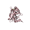
| ||||||||
| 4 | 
| ||||||||
| 5 | 
| ||||||||
| Unit cell |
|
-Components
-Yellowtail Ascites Virus (YAV) VP4 ... , 3 types, 5 molecules AEBCD
| #1: Protein | Mass: 22344.418 Da / Num. of mol.: 2 / Mutation: N616D Source method: isolated from a genetically manipulated source Source: (gene. exp.) Yellowtail ascites virus / Strain: Y-6 / Gene: VIRAL PROTEIN 4 (VP4) / Plasmid: PET28B+ / Production host:  #2: Protein | Mass: 22328.418 Da / Num. of mol.: 2 / Mutation: N616D Source method: isolated from a genetically manipulated source Source: (gene. exp.) Yellowtail ascites virus / Strain: Y-6 / Gene: VIRAL PROTEIN 4 (VP4) / Plasmid: PET28B+ / Production host: #3: Protein | | Mass: 22328.418 Da / Num. of mol.: 1 / Mutation: N616D Source method: isolated from a genetically manipulated source Source: (gene. exp.) Yellowtail ascites virus / Strain: Y-6 / Gene: VIRAL PROTEIN 4 (VP4) / Plasmid: PET28B+ / Production host: |
|---|
-Non-polymers , 4 types, 279 molecules 


| #4: Chemical | ChemComp-BME /  Mass: 78.133 Da / Num. of mol.: 6 / Source method: obtained synthetically / Formula: C2H6OS Mass: 78.133 Da / Num. of mol.: 6 / Source method: obtained synthetically / Formula: C2H6OS#5: Chemical | ChemComp-MG / |  Mass: 24.305 Da / Num. of mol.: 1 / Source method: obtained synthetically / Formula: Mg Mass: 24.305 Da / Num. of mol.: 1 / Source method: obtained synthetically / Formula: Mg#6: Chemical | ChemComp-GOL / |  Mass: 92.094 Da / Num. of mol.: 1 / Source method: obtained synthetically / Formula: C3H8O3 Mass: 92.094 Da / Num. of mol.: 1 / Source method: obtained synthetically / Formula: C3H8O3#7: Water | ChemComp-HOH / | Mass: 18.015 Da / Num. of mol.: 271 / Source method: isolated from a natural source / Formula: H2O |
|---|
-Experimental details
-Experiment
| Experiment | Method: X-RAY DIFFRACTION / Number of used crystals: 1 |
|---|
- Sample preparation
| Crystal | Density Matthews: 2.24 Å3/Da / Density % sol: 45.05 % |
|---|---|
| Crystal grow | Temperature: 296 K / Method: vapor diffusion, sitting drop / pH: 6.5 Details: 35% PEG2000, 0.3M Magnesium Chloride, 0.1M MES, pH 6.5, VAPOR DIFFUSION, SITTING DROP, temperature 296K |
-Data collection
| Diffraction | Mean temperature: 95 K |
|---|---|
| Diffraction source | Source: ROTATING ANODE / Type: RIGAKU MICROMAX-007 / Wavelength: 1.5418 Å |
| Detector | Type: RIGAKU RAXIS IV++ / Detector: IMAGE PLATE / Date: Aug 5, 2009 / Details: MULTILAYER OPTIC |
| Radiation | Monochromator: VARIMAX CU HF OPTICS / Protocol: SINGLE WAVELENGTH / Monochromatic (M) / Laue (L): M / Scattering type: x-ray |
| Radiation wavelength | Wavelength: 1.5418 Å / Relative weight: 1 |
| Reflection | Resolution: 2.5→64.3 Å / Num. all: 33009 / Num. obs: 33009 / % possible obs: 95.9 % / Observed criterion σ(F): 0 / Observed criterion σ(I): 0 / Redundancy: 3.7 % / Biso Wilson estimate: 40 Å2 / Rmerge(I) obs: 0.088 / Net I/σ(I): 10.5 |
| Reflection shell | Resolution: 2.5→2.6 Å / Redundancy: 3.3 % / Rmerge(I) obs: 0.278 / Mean I/σ(I) obs: 3.8 / Num. unique all: 4043 / % possible all: 80.7 |
- Processing
| Software |
| |||||||||||||||||||||||||||||||||||||||||||||
|---|---|---|---|---|---|---|---|---|---|---|---|---|---|---|---|---|---|---|---|---|---|---|---|---|---|---|---|---|---|---|---|---|---|---|---|---|---|---|---|---|---|---|---|---|---|---|
| Refinement | Method to determine structure: MOLECULAR REPLACEMENT Starting model: PDB entry 2PNM Resolution: 2.5→64.3 Å / Cor.coef. Fo:Fc: 0.943 / Cor.coef. Fo:Fc free: 0.903 / SU B: 9.852 / SU ML: 0.216 / Cross valid method: THROUGHOUT / σ(F): 0 / ESU R Free: 0.316 / Stereochemistry target values: MAXIMUM LIKELIHOOD / Details: HYDROGENS HAVE BEEN USED IF PRESENT IN THE INPUT
| |||||||||||||||||||||||||||||||||||||||||||||
| Solvent computation | Ion probe radii: 0.8 Å / Shrinkage radii: 0.8 Å / VDW probe radii: 1.2 Å / Solvent model: MASK | |||||||||||||||||||||||||||||||||||||||||||||
| Displacement parameters | Biso mean: 26.761 Å2
| |||||||||||||||||||||||||||||||||||||||||||||
| Refinement step | Cycle: LAST / Resolution: 2.5→64.3 Å
| |||||||||||||||||||||||||||||||||||||||||||||
| Refine LS restraints |
| |||||||||||||||||||||||||||||||||||||||||||||
| LS refinement shell | Resolution: 2.5→2.6 Å / Total num. of bins used: 20
|
