 Movie
Movie Controller


+ Open data

- Basic information
| Entry | Database: PDB / ID: 5v2v | ||||||
|---|---|---|---|---|---|---|---|
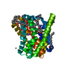
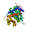
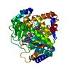
| Title | Ethylene forming enzyme in complex with nickel | ||||||
 Components Components | 2-oxoglutarate-dependent ethylene/succinate-forming enzyme | ||||||
 Keywords Keywords | OXIDOREDUCTASE / ethylene biosynthesis | ||||||
| Function / homology |  Function and homology information Function and homology information2-oxoglutarate dioxygenase (ethene-forming) / 2-oxoglutarate/L-arginine monooxygenase/decarboxylase (succinate-forming) / 2-oxoglutarate oxygenase/decarboxylase (ethylene-forming) activity / ethylene biosynthetic process / dioxygenase activity / metal ion binding Similarity search - Function | ||||||
| Biological species |  Pseudomonas savastanoi pv. phaseolicola (bacteria) Pseudomonas savastanoi pv. phaseolicola (bacteria) | ||||||
| Method |  X-RAY DIFFRACTION / SYNCHROTRON / MOLECULAR REPLACEMENT / molecular replacement / Resolution: 3.04 Å X-RAY DIFFRACTION / SYNCHROTRON / MOLECULAR REPLACEMENT / molecular replacement / Resolution: 3.04 Å | ||||||
 Authors Authors | Fellner, M. / Martinez, S. / Hu, J. / Hausinger, R.P. | ||||||
| Funding support |  United States, 1items United States, 1items
| ||||||
 Citation Citation | Journal: J. Am. Chem. Soc. / Year: 2017 Title: Structures and Mechanisms of the Non-Heme Fe(II)- and 2-Oxoglutarate-Dependent Ethylene-Forming Enzyme: Substrate Binding Creates a Twist. Authors: Martinez, S. / Fellner, M. / Herr, C.Q. / Ritchie, A. / Hu, J. / Hausinger, R.P. | ||||||
| History |
|
- Structure visualization
| Structure viewer | Molecule: MolmilJmol/JSmol |
|---|
- Downloads & links
-Download
| PDBx/mmCIF format | 5v2v.cif.gz | 74.6 KB | Display | PDBx/mmCIF format |
|---|---|---|---|---|
| PDB format | pdb5v2v.ent.gz | 52.6 KB | Display | PDB format |
| PDBx/mmJSON format | 5v2v.json.gz | Tree view | PDBx/mmJSON format | |
| Others |  Other downloads Other downloads |
-Validation report
| Arichive directory | https://data.pdbj.org/pub/pdb/validation_reports/v2/5v2vftp://data.pdbj.org/pub/pdb/validation_reports/v2/5v2v | HTTPS FTP |
|---|
-Related structure data
| Related structure data |  5v2tSC  5v2uC  5v2xC  5v2yC  5v2zC  5v31C  5v32C  5v34C  5vkaC  5vkbC S: Starting model for refinement C: citing same article ( |
|---|---|
| Similar structure data |


-Links
 PDBj
PDBj
- Assembly
| Deposited unit | 
| ||||||||
|---|---|---|---|---|---|---|---|---|---|
| 1 |
| ||||||||
| Unit cell |
|
-Components
| #1: Protein | Mass: 40138.809 Da / Num. of mol.: 1 Source method: isolated from a genetically manipulated source Source: (gene. exp.) Pseudomonas savastanoi pv. phaseolicola (bacteria)Gene: efe / Plasmid: pET28 / Production host: References: UniProt: P32021, 2-oxoglutarate dioxygenase (ethene-forming), EC: 1.14.11.34 |
|---|---|
| #2: Chemical | ChemComp-NI /   Mass: 58.693 Da / Num. of mol.: 1 / Source method: obtained synthetically / Formula: Ni Mass: 58.693 Da / Num. of mol.: 1 / Source method: obtained synthetically / Formula: Ni |
| #3: Water | ChemComp-HOH /  Mass: 18.015 Da / Num. of mol.: 2 / Source method: isolated from a natural source / Formula: H2O Mass: 18.015 Da / Num. of mol.: 2 / Source method: isolated from a natural source / Formula: H2O |
| Has protein modification | Y |
-Experimental details
-Experiment
| Experiment | Method: X-RAY DIFFRACTION / Number of used crystals: 1 |
|---|
- Sample preparation
| Crystal | Density Matthews: 1.81 Å3/Da / Density % sol: 32.1 % / Mosaicity: 1.57 ° |
|---|---|
| Crystal grow | Temperature: 277.15 K / Method: vapor diffusion, sitting drop / pH: 8 Details: 0.5 ul 72 mg/ml EFE (25 mM HEPES pH 8.0, 1 mM TCEP) was mixed with 0.5 ul reservoir solution. The sitting drop reservoir of 200 ul contained 0.2 M lithium chloride, 0.1 M Tris-HCl pH 8.0, ...Details: 0.5 ul 72 mg/ml EFE (25 mM HEPES pH 8.0, 1 mM TCEP) was mixed with 0.5 ul reservoir solution. The sitting drop reservoir of 200 ul contained 0.2 M lithium chloride, 0.1 M Tris-HCl pH 8.0, 20% w/v Polyethylene glycol 6,000. A grown crystal was soaked for 2 hours in a sitting drop containing 0.1 M nickel chloride, 0.2 M lithium chloride, 0.1 M Tris-HCl pH 8.0, 20% w/v Polyethylene glycol 6,000, the reservoir of 100 ul contained 0.2 M lithium chloride, 0.1 M Tris-HCl pH 8.0, 20% w/v Polyethylene glycol 6,000.The crystal was direcly frozen. |
-Data collection
| Diffraction | Mean temperature: 100 K | ||||||||||||||||||||||||
|---|---|---|---|---|---|---|---|---|---|---|---|---|---|---|---|---|---|---|---|---|---|---|---|---|---|
| Diffraction source | Source: SYNCHROTRON / Site: APS / Beamline: 21-ID-D / Wavelength: 1.4777 Å | ||||||||||||||||||||||||
| Detector | Type: DECTRIS EIGER X 9M / Detector: PIXEL / Date: Dec 14, 2016 | ||||||||||||||||||||||||
| Radiation | Protocol: SINGLE WAVELENGTH / Monochromatic (M) / Laue (L): M / Scattering type: x-ray | ||||||||||||||||||||||||
| Radiation wavelength | Wavelength: 1.4777 Å / Relative weight: 1 | ||||||||||||||||||||||||
| Reflection | Resolution: 3.04→87.75 Å / Num. obs: 5959 / % possible obs: 99.3 % / Redundancy: 4.8 % / Biso Wilson estimate: 54.66 Å2 / CC1/2: 0.99 / Rmerge(I) obs: 0.141 / Rpim(I) all: 0.069 / Rrim(I) all: 0.157 / Net I/σ(I): 7 / Num. measured all: 28385 / Scaling rejects: 32 | ||||||||||||||||||||||||
| Reflection shell | Diffraction-ID: 1
|
-Phasing
| Phasing | Method: molecular replacement | |||||||||
|---|---|---|---|---|---|---|---|---|---|---|
| Phasing MR |
|
- Processing
| Software |
| ||||||||||||||||||||||||||||||||||||||||||||||||||||||||||||||||||||||||||||||||||||
|---|---|---|---|---|---|---|---|---|---|---|---|---|---|---|---|---|---|---|---|---|---|---|---|---|---|---|---|---|---|---|---|---|---|---|---|---|---|---|---|---|---|---|---|---|---|---|---|---|---|---|---|---|---|---|---|---|---|---|---|---|---|---|---|---|---|---|---|---|---|---|---|---|---|---|---|---|---|---|---|---|---|---|---|---|---|
| Refinement | Method to determine structure: MOLECULAR REPLACEMENT Starting model: 5V2T Resolution: 3.04→43.875 Å / SU ML: 0.4 / Cross valid method: FREE R-VALUE / σ(F): 1.93 / Phase error: 28.96
| ||||||||||||||||||||||||||||||||||||||||||||||||||||||||||||||||||||||||||||||||||||
| Solvent computation | Shrinkage radii: 0.9 Å / VDW probe radii: 1.11 Å | ||||||||||||||||||||||||||||||||||||||||||||||||||||||||||||||||||||||||||||||||||||
| Displacement parameters | Biso max: 108.48 Å2 / Biso mean: 47.67 Å2 / Biso min: 23.31 Å2 | ||||||||||||||||||||||||||||||||||||||||||||||||||||||||||||||||||||||||||||||||||||
| Refinement step | Cycle: final / Resolution: 3.04→43.875 Å
| ||||||||||||||||||||||||||||||||||||||||||||||||||||||||||||||||||||||||||||||||||||
| Refine LS restraints |
| ||||||||||||||||||||||||||||||||||||||||||||||||||||||||||||||||||||||||||||||||||||
| LS refinement shell | Refine-ID: X-RAY DIFFRACTION / Rfactor Rfree error: 0 / Total num. of bins used: 11
|
