 Movie
Movie Controller

[English] 日本語
 Yorodumi
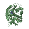
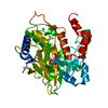
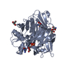
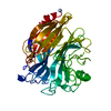
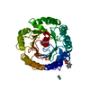
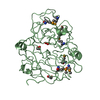
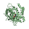
- PDB-5v34: Ethylene forming enzyme in complex with manganese, malic acid and... -
+ Open data

- Basic information
| Entry | Database: PDB / ID: 5v34 | ||||||
|---|---|---|---|---|---|---|---|
| Title | Ethylene forming enzyme in complex with manganese, malic acid and L-arginine | ||||||
 Components Components | 2-oxoglutarate-dependent ethylene/succinate-forming enzyme | ||||||
 Keywords Keywords | OXIDOREDUCTASE / ethylene biosynthesis | ||||||
| Function / homology |  Function and homology information Function and homology information2-oxoglutarate dioxygenase (ethene-forming) / 2-oxoglutarate/L-arginine monooxygenase/decarboxylase (succinate-forming) / 2-oxoglutarate oxygenase/decarboxylase (ethylene-forming) activity / ethylene biosynthetic process / dioxygenase activity / metal ion binding Similarity search - Function | ||||||
| Biological species |  Pseudomonas savastanoi pv. phaseolicola (bacteria) Pseudomonas savastanoi pv. phaseolicola (bacteria) | ||||||
| Method |  X-RAY DIFFRACTION / SYNCHROTRON / MOLECULAR REPLACEMENT / molecular replacement / Resolution: 1.48 Å X-RAY DIFFRACTION / SYNCHROTRON / MOLECULAR REPLACEMENT / molecular replacement / Resolution: 1.48 Å | ||||||
 Authors Authors | Fellner, M. / Martinez, S. / Hu, J. / Hausinger, R.P. | ||||||
| Funding support |  United States, 1items United States, 1items
| ||||||
 Citation Citation | Journal: J. Am. Chem. Soc. / Year: 2017 Title: Structures and Mechanisms of the Non-Heme Fe(II)- and 2-Oxoglutarate-Dependent Ethylene-Forming Enzyme: Substrate Binding Creates a Twist. Authors: Martinez, S. / Fellner, M. / Herr, C.Q. / Ritchie, A. / Hu, J. / Hausinger, R.P. | ||||||
| History |
|
- Structure visualization
| Structure viewer | Molecule: MolmilJmol/JSmol |
|---|
- Downloads & links
-Download
| PDBx/mmCIF format | 5v34.cif.gz | 223 KB | Display | PDBx/mmCIF format |
|---|---|---|---|---|
| PDB format | pdb5v34.ent.gz | 179.3 KB | Display | PDB format |
| PDBx/mmJSON format | 5v34.json.gz | Tree view | PDBx/mmJSON format | |
| Others |  Other downloads Other downloads |
-Validation report
| Arichive directory | https://data.pdbj.org/pub/pdb/validation_reports/v3/5v34ftp://data.pdbj.org/pub/pdb/validation_reports/v3/5v34 | HTTPS FTP |
|---|
-Related structure data
| Related structure data |  5v2tSC  5v2uC  5v2vC  5v2xC  5v2yC  5v2zC  5v31C  5v32C  5vkaC  5vkbC S: Starting model for refinement C: citing same article ( |
|---|---|
| Similar structure data |


-Links
 PDBj
PDBj


- Assembly
| Deposited unit | 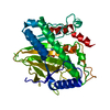
| ||||||||||||
|---|---|---|---|---|---|---|---|---|---|---|---|---|---|
| 1 |
| ||||||||||||
| Unit cell |
| ||||||||||||
| Components on special symmetry positions |
|
-Components
| #1: Protein | Mass: 39716.762 Da / Num. of mol.: 1 Source method: isolated from a genetically manipulated source Source: (gene. exp.) Pseudomonas savastanoi pv. phaseolicola (bacteria)Gene: efe / Plasmid: pET28 / Production host: References: UniProt: P32021, 2-oxoglutarate dioxygenase (ethene-forming), EC: 1.14.11.34 |
|---|---|
| #2: Chemical | ChemComp-ARG /   Type: L-peptide linking / Mass: 175.209 Da / Num. of mol.: 1 / Source method: obtained synthetically / Formula: C6H15N4O2 Type: L-peptide linking / Mass: 175.209 Da / Num. of mol.: 1 / Source method: obtained synthetically / Formula: C6H15N4O2 |
| #3: Chemical | ChemComp-MN /   Mass: 54.938 Da / Num. of mol.: 1 / Source method: obtained synthetically / Formula: Mn Mass: 54.938 Da / Num. of mol.: 1 / Source method: obtained synthetically / Formula: Mn |
| #4: Chemical | ChemComp-MLT /   Mass: 134.087 Da / Num. of mol.: 1 / Source method: obtained synthetically / Formula: C4H6O5 Mass: 134.087 Da / Num. of mol.: 1 / Source method: obtained synthetically / Formula: C4H6O5 |
| #5: Water | ChemComp-HOH /  Mass: 18.015 Da / Num. of mol.: 473 / Source method: isolated from a natural source / Formula: H2O Mass: 18.015 Da / Num. of mol.: 473 / Source method: isolated from a natural source / Formula: H2O |
-Experimental details
-Experiment
| Experiment | Method: X-RAY DIFFRACTION / Number of used crystals: 1 |
|---|
- Sample preparation
| Crystal | Density Matthews: 2.89 Å3/Da / Density % sol: 57.48 % / Mosaicity: 0.38 ° |
|---|---|
| Crystal grow | Temperature: 277.15 K / Method: vapor diffusion, sitting drop / pH: 6.5 Details: 0.2 ul 64 mg/ml EFE (+1 mM manganese chloride, 25 mM HEPES pH 8.0, 1 mM TCEP, +2.5 mM L-arginine) was mixed with 0.2 ul reservoir solution. The sitting drop reservoir of 50 ul contained 25% ...Details: 0.2 ul 64 mg/ml EFE (+1 mM manganese chloride, 25 mM HEPES pH 8.0, 1 mM TCEP, +2.5 mM L-arginine) was mixed with 0.2 ul reservoir solution. The sitting drop reservoir of 50 ul contained 25% PEG 1500 and 0.1 M MMT buffer, pH 6.5. The crystal was soaked for about a minute in 25% w/v Polyethylene glycol 400, 75% reservoir solution before freezing it. |
-Data collection
| Diffraction | Mean temperature: 100 K | ||||||||||||||||||||||||
|---|---|---|---|---|---|---|---|---|---|---|---|---|---|---|---|---|---|---|---|---|---|---|---|---|---|
| Diffraction source | Source: SYNCHROTRON / Site: APS / Beamline: 21-ID-D / Wavelength: 1.1272 Å | ||||||||||||||||||||||||
| Detector | Type: DECTRIS EIGER X 9M / Detector: PIXEL / Date: Feb 11, 2017 | ||||||||||||||||||||||||
| Radiation | Protocol: SINGLE WAVELENGTH / Monochromatic (M) / Laue (L): M / Scattering type: x-ray | ||||||||||||||||||||||||
| Radiation wavelength | Wavelength: 1.1272 Å / Relative weight: 1 | ||||||||||||||||||||||||
| Reflection | Resolution: 1.48→61.24 Å / Num. obs: 75179 / % possible obs: 97.9 % / Redundancy: 7.2 % / Biso Wilson estimate: 16.94 Å2 / CC1/2: 0.998 / Rmerge(I) obs: 0.084 / Rpim(I) all: 0.033 / Rrim(I) all: 0.091 / Net I/σ(I): 12.8 | ||||||||||||||||||||||||
| Reflection shell | Diffraction-ID: 1
|
-Phasing
| Phasing | Method: molecular replacement | |||||||||
|---|---|---|---|---|---|---|---|---|---|---|
| Phasing MR |
|
- Processing
| Software |
| |||||||||||||||||||||||||||||||||||||||||||||||||||||||||||||||||||||||||||||||||||||||||||||||||||||||||||||||||||||||||||||||||||||||||||||||||||||||||||||||||||||||||||||||||||||||||||||||||||||||||||||||||||||||||
|---|---|---|---|---|---|---|---|---|---|---|---|---|---|---|---|---|---|---|---|---|---|---|---|---|---|---|---|---|---|---|---|---|---|---|---|---|---|---|---|---|---|---|---|---|---|---|---|---|---|---|---|---|---|---|---|---|---|---|---|---|---|---|---|---|---|---|---|---|---|---|---|---|---|---|---|---|---|---|---|---|---|---|---|---|---|---|---|---|---|---|---|---|---|---|---|---|---|---|---|---|---|---|---|---|---|---|---|---|---|---|---|---|---|---|---|---|---|---|---|---|---|---|---|---|---|---|---|---|---|---|---|---|---|---|---|---|---|---|---|---|---|---|---|---|---|---|---|---|---|---|---|---|---|---|---|---|---|---|---|---|---|---|---|---|---|---|---|---|---|---|---|---|---|---|---|---|---|---|---|---|---|---|---|---|---|---|---|---|---|---|---|---|---|---|---|---|---|---|---|---|---|---|---|---|---|---|---|---|---|---|---|---|---|---|---|---|---|---|
| Refinement | Method to determine structure: MOLECULAR REPLACEMENT Starting model: 5V2T Resolution: 1.48→43.68 Å / SU ML: 0.17 / Cross valid method: FREE R-VALUE / σ(F): 1.92 / Phase error: 15.51
| |||||||||||||||||||||||||||||||||||||||||||||||||||||||||||||||||||||||||||||||||||||||||||||||||||||||||||||||||||||||||||||||||||||||||||||||||||||||||||||||||||||||||||||||||||||||||||||||||||||||||||||||||||||||||
| Solvent computation | Shrinkage radii: 0.9 Å / VDW probe radii: 1.11 Å | |||||||||||||||||||||||||||||||||||||||||||||||||||||||||||||||||||||||||||||||||||||||||||||||||||||||||||||||||||||||||||||||||||||||||||||||||||||||||||||||||||||||||||||||||||||||||||||||||||||||||||||||||||||||||
| Displacement parameters | Biso max: 73.51 Å2 / Biso mean: 23.777 Å2 / Biso min: 10.86 Å2 | |||||||||||||||||||||||||||||||||||||||||||||||||||||||||||||||||||||||||||||||||||||||||||||||||||||||||||||||||||||||||||||||||||||||||||||||||||||||||||||||||||||||||||||||||||||||||||||||||||||||||||||||||||||||||
| Refinement step | Cycle: final / Resolution: 1.48→43.68 Å
| |||||||||||||||||||||||||||||||||||||||||||||||||||||||||||||||||||||||||||||||||||||||||||||||||||||||||||||||||||||||||||||||||||||||||||||||||||||||||||||||||||||||||||||||||||||||||||||||||||||||||||||||||||||||||
| Refine LS restraints |
| |||||||||||||||||||||||||||||||||||||||||||||||||||||||||||||||||||||||||||||||||||||||||||||||||||||||||||||||||||||||||||||||||||||||||||||||||||||||||||||||||||||||||||||||||||||||||||||||||||||||||||||||||||||||||
| LS refinement shell | Refine-ID: X-RAY DIFFRACTION / Rfactor Rfree error: 0 / Total num. of bins used: 30
|
