 Movie
Movie Controller

[English] 日本語
 Yorodumi
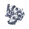
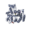
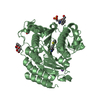
- PDB-3f0r: Crystal Structure Analysis of Human HDAC8 complexed with trichost... -
+ Open data

- Basic information
| Entry | Database: PDB / ID: 3f0r | ||||||
|---|---|---|---|---|---|---|---|
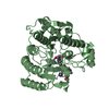
| Title | Crystal Structure Analysis of Human HDAC8 complexed with trichostatin A in a new monoclinic crystal form | ||||||
 Components Components | Histone deacetylase 8 | ||||||
 Keywords Keywords | HYDROLASE / HDAC / metalloenzyme / acetylation / arginase fold / HDAC8 / histone deacetylase 8 / hydroxamate inhibitor / Alternative splicing / Chromatin regulator / Nucleus / Repressor / Transcription / Transcription regulation | ||||||
| Function / homology |  Function and homology information Function and homology informationhistone decrotonylase activity / histone deacetylase activity, hydrolytic mechanism / histone deacetylase / histone deacetylase activity / protein lysine deacetylase activity / Hydrolases; Acting on carbon-nitrogen bonds, other than peptide bonds; In linear amides / regulation of telomere maintenance / mitotic sister chromatid cohesion / Notch-HLH transcription pathway / nuclear chromosome ...histone decrotonylase activity / histone deacetylase activity, hydrolytic mechanism / histone deacetylase / histone deacetylase activity / protein lysine deacetylase activity / Hydrolases; Acting on carbon-nitrogen bonds, other than peptide bonds; In linear amides / regulation of telomere maintenance / mitotic sister chromatid cohesion / Notch-HLH transcription pathway / nuclear chromosome / histone deacetylase complex / negative regulation of protein ubiquitination / Hsp70 protein binding / Resolution of Sister Chromatid Cohesion / HDACs deacetylate histones / regulation of protein stability / Hsp90 protein binding / NOTCH1 Intracellular Domain Regulates Transcription / Constitutive Signaling by NOTCH1 PEST Domain Mutants / Constitutive Signaling by NOTCH1 HD+PEST Domain Mutants / Separation of Sister Chromatids / heterochromatin formation / chromatin organization / DNA-binding transcription factor binding / negative regulation of transcription by RNA polymerase II / nucleoplasm / metal ion binding / nucleus / cytoplasm Similarity search - Function | ||||||
| Biological species |  Homo sapiens (human) Homo sapiens (human) | ||||||
| Method |  X-RAY DIFFRACTION / SYNCHROTRON / MOLECULAR REPLACEMENT / Resolution: 2.54 Å X-RAY DIFFRACTION / SYNCHROTRON / MOLECULAR REPLACEMENT / Resolution: 2.54 Å | ||||||
 Authors Authors | Dowling, D.P. / Gantt, S.L. / Gattis, S.G. / Fierke, C.A. / Christianson, D.W. | ||||||
 Citation Citation | Journal: Biochemistry / Year: 2008 Title: Structural studies of human histone deacetylase 8 and its site-specific variants complexed with substrate and inhibitors. Authors: Dowling, D.P. / Gantt, S.L. / Gattis, S.G. / Fierke, C.A. / Christianson, D.W. | ||||||
| History |
|
- Structure visualization
| Structure viewer | Molecule: MolmilJmol/JSmol |
|---|
- Downloads & links
-Download
| PDBx/mmCIF format | 3f0r.cif.gz | 224.6 KB | Display | PDBx/mmCIF format |
|---|---|---|---|---|
| PDB format | pdb3f0r.ent.gz | 181.2 KB | Display | PDB format |
| PDBx/mmJSON format | 3f0r.json.gz | Tree view | PDBx/mmJSON format | |
| Others |  Other downloads Other downloads |
-Validation report
| Arichive directory | https://data.pdbj.org/pub/pdb/validation_reports/f0/3f0rftp://data.pdbj.org/pub/pdb/validation_reports/f0/3f0r | HTTPS FTP |
|---|
-Related structure data
| Related structure data |  3ew8C  3ewfC  3ezpC  3eztC 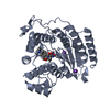 3f06C  3f07C  1w22S C: citing same article ( S: Starting model for refinement |
|---|---|
| Similar structure data |


-Links
 PDBj
PDBj


- Assembly
| Deposited unit | 
| ||||||||
|---|---|---|---|---|---|---|---|---|---|
| 1 | 
| ||||||||
| 2 | 
| ||||||||
| 3 | 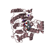
| ||||||||
| Unit cell |
|
-Components
| #1: Protein | Mass: 43174.941 Da / Num. of mol.: 3 Source method: isolated from a genetically manipulated source Source: (gene. exp.) Homo sapiens (human) / Gene: CDA07, HDAC8, HDACL1 / Plasmid: pHD2-Xa-His / Production host:  #2: Chemical |   Mass: 65.409 Da / Num. of mol.: 3 / Source method: obtained synthetically / Formula: Zn Mass: 65.409 Da / Num. of mol.: 3 / Source method: obtained synthetically / Formula: Zn#3: Chemical | ChemComp-K /   Mass: 39.098 Da / Num. of mol.: 6 / Source method: obtained synthetically / Formula: K Mass: 39.098 Da / Num. of mol.: 6 / Source method: obtained synthetically / Formula: K#4: Chemical | ChemComp-TSN /   Mass: 302.368 Da / Num. of mol.: 6 / Source method: obtained synthetically / Formula: C17H22N2O3 Mass: 302.368 Da / Num. of mol.: 6 / Source method: obtained synthetically / Formula: C17H22N2O3#5: Water | ChemComp-HOH / |  Mass: 18.015 Da / Num. of mol.: 101 / Source method: isolated from a natural source / Formula: H2O Mass: 18.015 Da / Num. of mol.: 101 / Source method: isolated from a natural source / Formula: H2O |
|---|
-Experimental details
-Experiment
| Experiment | Method: X-RAY DIFFRACTION / Number of used crystals: 1 |
|---|
- Sample preparation
| Crystal | Density Matthews: 2.83 Å3/Da / Density % sol: 56.48 % |
|---|---|
| Crystal grow | Temperature: 298 K / Method: vapor diffusion, sitting drop / pH: 5.8 Details: Final drop concentrations of 25 mM Tris, 2.5% glycerol, 75 mM KCl, 1-5% PEG 6000, 50 mM MES, 1 mM tri(2-carboxyethyl)phosphine (TCEP), 0.03 mM gly-gly-gly , pH 5.8, VAPOR DIFFUSION, SITTING ...Details: Final drop concentrations of 25 mM Tris, 2.5% glycerol, 75 mM KCl, 1-5% PEG 6000, 50 mM MES, 1 mM tri(2-carboxyethyl)phosphine (TCEP), 0.03 mM gly-gly-gly , pH 5.8, VAPOR DIFFUSION, SITTING DROP, temperature 298K |
-Data collection
| Diffraction | Mean temperature: 100 K |
|---|---|
| Diffraction source | Source: SYNCHROTRON / Site: CHESS  / Beamline: F1 / Wavelength: 0.9124 Å / Beamline: F1 / Wavelength: 0.9124 Å |
| Detector | Type: ADSC QUANTUM 270 / Detector: CCD / Date: Feb 3, 2006 |
| Radiation | Protocol: SINGLE WAVELENGTH / Monochromatic (M) / Laue (L): M / Scattering type: x-ray |
| Radiation wavelength | Wavelength: 0.9124 Å / Relative weight: 1 |
| Reflection | Resolution: 2.54→50 Å / Num. all: 45639 / Num. obs: 45639 / % possible obs: 96.8 % / Redundancy: 3.2 % / Biso Wilson estimate: 63.4 Å2 / Rmerge(I) obs: 0.097 / Net I/σ(I): 11.7 |
| Reflection shell | Resolution: 2.54→2.63 Å / Redundancy: 3 % / Rmerge(I) obs: 0.508 / Mean I/σ(I) obs: 2.3 / Num. unique all: 4566 / % possible all: 97.4 |
- Processing
| Software |
| ||||||||||||||||||||||||||||||||||||||||||||||||||||||||||||||||||||||||||||||||
|---|---|---|---|---|---|---|---|---|---|---|---|---|---|---|---|---|---|---|---|---|---|---|---|---|---|---|---|---|---|---|---|---|---|---|---|---|---|---|---|---|---|---|---|---|---|---|---|---|---|---|---|---|---|---|---|---|---|---|---|---|---|---|---|---|---|---|---|---|---|---|---|---|---|---|---|---|---|---|---|---|---|
| Refinement | Method to determine structure: MOLECULAR REPLACEMENT Starting model: PDB entry 1W22 Resolution: 2.54→40.97 Å / Rfactor Rfree error: 0.006 / Data cutoff high absF: 87356.13 / Data cutoff low absF: 0 / Isotropic thermal model: RESTRAINED / Cross valid method: THROUGHOUT / σ(F): 0 / Stereochemistry target values: Engh & Huber / Details: BULK SOLVENT MODEL USED
| ||||||||||||||||||||||||||||||||||||||||||||||||||||||||||||||||||||||||||||||||
| Solvent computation | Solvent model: FLAT MODEL / Bsol: 43.55 Å2 / ksol: 0.35 e/Å3 | ||||||||||||||||||||||||||||||||||||||||||||||||||||||||||||||||||||||||||||||||
| Displacement parameters | Biso mean: 57.9 Å2
| ||||||||||||||||||||||||||||||||||||||||||||||||||||||||||||||||||||||||||||||||
| Refine analyze |
| ||||||||||||||||||||||||||||||||||||||||||||||||||||||||||||||||||||||||||||||||
| Refinement step | Cycle: LAST / Resolution: 2.54→40.97 Å
| ||||||||||||||||||||||||||||||||||||||||||||||||||||||||||||||||||||||||||||||||
| Refine LS restraints |
| ||||||||||||||||||||||||||||||||||||||||||||||||||||||||||||||||||||||||||||||||
| LS refinement shell | Resolution: 2.54→2.7 Å / Rfactor Rfree error: 0.02 / Total num. of bins used: 6
| ||||||||||||||||||||||||||||||||||||||||||||||||||||||||||||||||||||||||||||||||
| Xplor file |
|
