- PDB-5ukw: Crystal structure of human Glucose 6-phosphate Dehydrogenase muta... -
+
Open data
ID or keywords:
Loading...
-
Basic information
Entry
Database: PDB / ID: 5ukw
Title
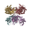
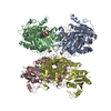
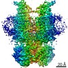
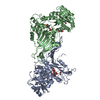
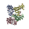
Crystal structure of human Glucose 6-phosphate Dehydrogenase mutant (A277C) complexed with G6P
Components
Glucose-6-phosphate 1-dehydrogenase
Keywords
OXIDOREDUCTASE / Dehydrogenase / A277C / Tetramer
Function / homology
Function and homology information
pentose biosynthetic process / ribose phosphate biosynthetic process / response to iron(III) ion / positive regulation of calcium ion transmembrane transport via high voltage-gated calcium channel / glucose-6-phosphate dehydrogenase (NADP+) / glucose-6-phosphate dehydrogenase activity / Pentose phosphate pathway / pentose-phosphate shunt, oxidative branch / negative regulation of cell growth involved in cardiac muscle cell development / glucose 6-phosphate metabolic process ...pentose biosynthetic process / ribose phosphate biosynthetic process / response to iron(III) ion / positive regulation of calcium ion transmembrane transport via high voltage-gated calcium channel / glucose-6-phosphate dehydrogenase (NADP+) / glucose-6-phosphate dehydrogenase activity / Pentose phosphate pathway / pentose-phosphate shunt, oxidative branch / negative regulation of cell growth involved in cardiac muscle cell development / glucose 6-phosphate metabolic process / NADP+ metabolic process / pentose-phosphate shunt / D-glucose binding / NFE2L2 regulates pentose phosphate pathway genes / Oxidoreductases / erythrocyte maturation / cholesterol biosynthetic process / response to food / negative regulation of reactive oxygen species metabolic process / substantia nigra development / regulation of neuron apoptotic process / TP53 Regulates Metabolic Genes / lipid metabolic process / glutathione metabolic process / glucose metabolic process / cytoplasmic side of plasma membrane / centriolar satellite / NADP binding / cellular response to oxidative stress / response to ethanol / protein homodimerization activity / extracellular exosome / membrane / identical protein binding / cytoplasm / cytosol Similarity search - Function
 Movie
Movie Controller

 Yorodumi
 Open data
 Basic information
 Components
 Keywords
 Function and homology information
 Homo sapiens (human)
 X-RAY DIFFRACTION /
  Authors
 Brazil, 1items
  Citation
 Structure visualization
 Downloads & links
 Other downloads
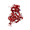


 PDBj
PDBj Assembly
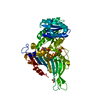

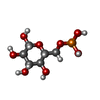
 Type: D-saccharide, beta linking / Mass: 260.136 Da / Num. of mol.: 1
 Mass: 92.094 Da / Num. of mol.: 1 / Source method: obtained synthetically / Formula: C3H8O3
 Mass: 18.015 Da / Num. of mol.: 20 / Source method: isolated from a natural source / Formula: H2O
 Sample preparation
 / Beamline: X06DA / Wavelength: 0.97643 Å
 Processing