 Movie
Movie Controller

[English] 日本語
 Yorodumi
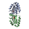
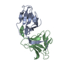
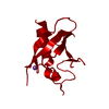
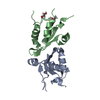
- PDB-5uff: Crystal Structure of Variable Lymphocyte Receptor (VLR) RBC36 wit... -
+ Open data

- Basic information
| Entry | Database: PDB / ID: 5uff | |||||||||
|---|---|---|---|---|---|---|---|---|---|---|
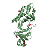
| Title | Crystal Structure of Variable Lymphocyte Receptor (VLR) RBC36 with Fucose(alpha-1-2)Lactose bound | |||||||||
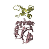
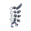
 Components Components | RBC36 | |||||||||
 Keywords Keywords | IMMUNE SYSTEM / variable lymphocyte receptors / VLR / leucine-rich repeat / LRR / adaptive immunity / sea lamprey / jawless fish / receptor / glycan binding / glycan receptor | |||||||||
| Function / homology | 2'-fucosyllactose Function and homology information Function and homology information | |||||||||
| Biological species |  | |||||||||
| Method |  X-RAY DIFFRACTION / SYNCHROTRON / MOLECULAR REPLACEMENT / Resolution: 2.137 Å X-RAY DIFFRACTION / SYNCHROTRON / MOLECULAR REPLACEMENT / Resolution: 2.137 Å | |||||||||
 Authors Authors | Collins, B.C. / Gunn, R.J. / McKitrick, T.R. / Herrin, B.R. / Cummings, R.D. / Cooper, M.D. / Wilson, I.A. | |||||||||
| Funding support |  United States, 1items United States, 1items
| |||||||||
 Citation Citation | Journal: Structure / Year: 2017 Title: Structural Insights into VLR Fine Specificity for Blood Group Carbohydrates. Authors: Collins, B.C. / Gunn, R.J. / McKitrick, T.R. / Cummings, R.D. / Cooper, M.D. / Herrin, B.R. / Wilson, I.A. | |||||||||
| History |
|
- Structure visualization
| Structure viewer | Molecule: MolmilJmol/JSmol |
|---|
- Downloads & links
-Download
| PDBx/mmCIF format | 5uff.cif.gz | 58.8 KB | Display | PDBx/mmCIF format |
|---|---|---|---|---|
| PDB format | pdb5uff.ent.gz | 39.3 KB | Display | PDB format |
| PDBx/mmJSON format | 5uff.json.gz | Tree view | PDBx/mmJSON format | |
| Others |  Other downloads Other downloads |
-Validation report
| Summary document | 5uff_validation.pdf.gz | 822.4 KB | Display | wwPDB validaton report |
|---|---|---|---|---|
| Full document | 5uff_full_validation.pdf.gz | 824.7 KB | Display | |
| Data in XML | 5uff_validation.xml.gz | 11 KB | Display | |
| Data in CIF | 5uff_validation.cif.gz | 14.3 KB | Display | |
| Arichive directory | https://data.pdbj.org/pub/pdb/validation_reports/uf/5uffftp://data.pdbj.org/pub/pdb/validation_reports/uf/5uff | HTTPS FTP |
-Related structure data
| Related structure data |  5ueiC  5uf1C  5uf4C  5ufbC  5ufcC  5ufdC  3e6jS C: citing same article ( S: Starting model for refinement |
|---|---|
| Similar structure data |


-Links
 PDBj
PDBj- Assembly
| Deposited unit | 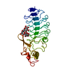
| ||||||||
|---|---|---|---|---|---|---|---|---|---|
| 1 |
| ||||||||
| Unit cell |
|
-Components
| #1: Protein | Mass: 23763.137 Da / Num. of mol.: 1 Source method: isolated from a genetically manipulated source Source: (gene. exp.)   Spodoptera frugiperda (fall armyworm) Spodoptera frugiperda (fall armyworm) |
|---|---|
| #2: Polysaccharide | alpha-L-fucopyranose-(1-2)-beta-D-galactopyranose-(1-4)-beta-D-glucopyranose / 2'-fucosyllactose  Source method: isolated from a genetically manipulated source Details: oligosaccharide / References: 2'-fucosyllactose |
| #3: Water | ChemComp-HOH /  Mass: 18.015 Da / Num. of mol.: 61 / Source method: isolated from a natural source / Formula: H2O Mass: 18.015 Da / Num. of mol.: 61 / Source method: isolated from a natural source / Formula: H2O |
| Has protein modification | Y |
-Experimental details
-Experiment
| Experiment | Method: X-RAY DIFFRACTION / Number of used crystals: 1 |
|---|
- Sample preparation
| Crystal | Density Matthews: 2.28 Å3/Da / Density % sol: 46.09 % |
|---|---|
| Crystal grow | Temperature: 277 K / Method: vapor diffusion, sitting drop Details: 11.4 mg/mL RBC36, 6.9 mM fucose(alpha-1-2)lactose, 140 mM ammonium chloride, 22% PEG3350 |
-Data collection
| Diffraction | Mean temperature: 100 K |
|---|---|
| Diffraction source | Source: SYNCHROTRON / Site: SSRL / Beamline: BL12-2 / Wavelength: 0.9784 Å |
| Detector | Type: DECTRIS PILATUS 6M / Detector: PIXEL / Date: May 5, 2015 |
| Radiation | Monochromator: Si(111) / Protocol: SINGLE WAVELENGTH / Monochromatic (M) / Laue (L): M / Scattering type: x-ray |
| Radiation wavelength | Wavelength: 0.9784 Å / Relative weight: 1 |
| Reflection | Resolution: 2.137→39.865 Å / Num. obs: 11758 / % possible obs: 97 % / Redundancy: 6.5 % / Rpim(I) all: 0.07 / Net I/σ(I): 8 |
| Reflection shell | Resolution: 2.137→2.21 Å / Num. measured obs: 7003 / Num. unique all: 1106 / Rpim(I) all: 0.7 |
- Processing
| Software |
| |||||||||||||||||||||||||||||||||||
|---|---|---|---|---|---|---|---|---|---|---|---|---|---|---|---|---|---|---|---|---|---|---|---|---|---|---|---|---|---|---|---|---|---|---|---|---|
| Refinement | Method to determine structure: MOLECULAR REPLACEMENT Starting model: PDB entry 3E6J Resolution: 2.137→39.856 Å / SU ML: 0.31 / Cross valid method: FREE R-VALUE / σ(F): 1.36 / Phase error: 39.58
| |||||||||||||||||||||||||||||||||||
| Solvent computation | Shrinkage radii: 0.9 Å / VDW probe radii: 1.11 Å | |||||||||||||||||||||||||||||||||||
| Displacement parameters | Biso max: 83.69 Å2 / Biso mean: 37.6366 Å2 / Biso min: 21.08 Å2 | |||||||||||||||||||||||||||||||||||
| Refinement step | Cycle: final / Resolution: 2.137→39.856 Å
| |||||||||||||||||||||||||||||||||||
| Refine LS restraints |
| |||||||||||||||||||||||||||||||||||
| LS refinement shell | Refine-ID: X-RAY DIFFRACTION / Rfactor Rfree error: 0 / Total num. of bins used: 4
|
