 Movie
Movie Controller


+ Open data

- Basic information
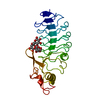
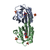
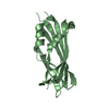
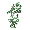
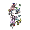
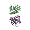
| Entry | Database: PDB / ID: 5uaz | ||||||
|---|---|---|---|---|---|---|---|
| Title | Crystal structure of the yeast nucleoporin | ||||||
 Components Components | Nucleoporin NUP53 | ||||||
 Keywords Keywords | PROTEIN TRANSPORT / nucleoporin / Nuclear Pore Complex / nucleocytoplasmic transport / dimerization domain | ||||||
| Function / homology |  Function and homology information Function and homology informationresponse to spindle checkpoint signaling / regulation of protein desumoylation / nuclear pore central transport channel / nuclear pore organization / nuclear pore nuclear basket / protein localization to kinetochore / structural constituent of nuclear pore / nucleocytoplasmic transport / NLS-bearing protein import into nucleus / regulation of mitotic nuclear division ...response to spindle checkpoint signaling / regulation of protein desumoylation / nuclear pore central transport channel / nuclear pore organization / nuclear pore nuclear basket / protein localization to kinetochore / structural constituent of nuclear pore / nucleocytoplasmic transport / NLS-bearing protein import into nucleus / regulation of mitotic nuclear division / nuclear pore / mRNA transport / phospholipid binding / protein import into nucleus / nuclear envelope / single-stranded DNA binding / nuclear membrane / cell division / positive regulation of DNA-templated transcription / identical protein binding Similarity search - Function | ||||||
| Biological species |  | ||||||
| Method |  X-RAY DIFFRACTION / SYNCHROTRON / MOLECULAR REPLACEMENT / Resolution: 1.753 Å X-RAY DIFFRACTION / SYNCHROTRON / MOLECULAR REPLACEMENT / Resolution: 1.753 Å | ||||||
 Authors Authors | Blus, B.J. / Blobel, G. | ||||||
| Funding support |  United States, 1items United States, 1items
| ||||||
 Citation Citation | Journal: To Be Published Title: Crystal structure of the yeast nucleoporin Authors: Blus, B.J. / Blobel, G. | ||||||
| History |
|
- Structure visualization
| Structure viewer | Molecule: MolmilJmol/JSmol |
|---|
- Downloads & links
-Download
| PDBx/mmCIF format | 5uaz.cif.gz | 126.8 KB | Display | PDBx/mmCIF format |
|---|---|---|---|---|
| PDB format | pdb5uaz.ent.gz | 100 KB | Display | PDB format |
| PDBx/mmJSON format | 5uaz.json.gz | Tree view | PDBx/mmJSON format | |
| Others |  Other downloads Other downloads |
-Validation report
| Arichive directory | https://data.pdbj.org/pub/pdb/validation_reports/ua/5uazftp://data.pdbj.org/pub/pdb/validation_reports/ua/5uaz | HTTPS FTP |
|---|
-Related structure data
| Related structure data |  3p3dS S: Starting model for refinement |
|---|---|
| Similar structure data |


-Links
 PDBj
PDBj
- Assembly
| Deposited unit | 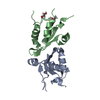
| |||||||||||||||
|---|---|---|---|---|---|---|---|---|---|---|---|---|---|---|---|---|
| 1 |
| |||||||||||||||
| Unit cell |
| |||||||||||||||
| Components on special symmetry positions |
|
-Components
| #1: Protein | Mass: 12419.255 Da / Num. of mol.: 2 Source method: isolated from a genetically manipulated source Source: (gene. exp.) Strain: ATCC 204508 / S288c / Gene: NUP53, YMR153W, YM8520.02 / Production host:  #2: Chemical |   Mass: 62.068 Da / Num. of mol.: 2 Mass: 62.068 Da / Num. of mol.: 2Source method: isolated from a genetically manipulated source Formula: C2H6O2 #3: Water | ChemComp-HOH / |  Mass: 18.015 Da / Num. of mol.: 135 / Source method: isolated from a natural source / Formula: H2O Mass: 18.015 Da / Num. of mol.: 135 / Source method: isolated from a natural source / Formula: H2O |
|---|
-Experimental details
-Experiment
| Experiment | Method: X-RAY DIFFRACTION / Number of used crystals: 1 |
|---|
- Sample preparation
| Crystal | Density Matthews: 1.79 Å3/Da / Density % sol: 31.23 % |
|---|---|
| Crystal grow | Temperature: 277 K / Method: vapor diffusion, sitting drop / pH: 7.5 / Details: 20% (wt/vol) PEG 3350 0.2 M Ammonium sulfate |
-Data collection
| Diffraction | Mean temperature: 100 K / Ambient temp details: nitrogen cryo-stream |
|---|---|
| Diffraction source | Source: SYNCHROTRON / Site: ALS / Beamline: 8.2.1 / Wavelength: 1 Å |
| Detector | Type: ADSC QUANTUM 315 / Detector: CCD / Date: May 22, 2016 |
| Radiation | Monochromator: Double-crystal Si(111) and multilayer / Protocol: SINGLE WAVELENGTH / Monochromatic (M) / Laue (L): M / Scattering type: x-ray |
| Radiation wavelength | Wavelength: 1 Å / Relative weight: 1 |
| Reflection | Resolution: 1.753→43.282 Å / Num. obs: 18275 / % possible obs: 99.85 % / Redundancy: 13.3 % / Biso Wilson estimate: 25.4 Å2 / Rmerge(I) obs: 0.119 / Rsym value: 0.119 / Net I/σ(I): 21.66 |
| Reflection shell | Resolution: 1.753→1.816 Å / Redundancy: 9.9 % / Mean I/σ(I) obs: 2.55 / CC1/2: 0.832 / % possible all: 98.72 |
- Processing
| Software |
| ||||||||||||||||||||||||||||||||||||||||||||||||||||||||||||||||||||||||||||||||||||||||||||||||||||||||||||||||||||||||||||||||||||||||||||||||||||||||||||||||||||||||||||||||||||||||||||||||||||||||||||||||||||||||||||||||||||||||||||||||||||||||||
|---|---|---|---|---|---|---|---|---|---|---|---|---|---|---|---|---|---|---|---|---|---|---|---|---|---|---|---|---|---|---|---|---|---|---|---|---|---|---|---|---|---|---|---|---|---|---|---|---|---|---|---|---|---|---|---|---|---|---|---|---|---|---|---|---|---|---|---|---|---|---|---|---|---|---|---|---|---|---|---|---|---|---|---|---|---|---|---|---|---|---|---|---|---|---|---|---|---|---|---|---|---|---|---|---|---|---|---|---|---|---|---|---|---|---|---|---|---|---|---|---|---|---|---|---|---|---|---|---|---|---|---|---|---|---|---|---|---|---|---|---|---|---|---|---|---|---|---|---|---|---|---|---|---|---|---|---|---|---|---|---|---|---|---|---|---|---|---|---|---|---|---|---|---|---|---|---|---|---|---|---|---|---|---|---|---|---|---|---|---|---|---|---|---|---|---|---|---|---|---|---|---|---|---|---|---|---|---|---|---|---|---|---|---|---|---|---|---|---|---|---|---|---|---|---|---|---|---|---|---|---|---|---|---|---|---|---|---|---|---|---|---|---|---|---|---|---|---|---|---|---|---|
| Refinement | Method to determine structure: MOLECULAR REPLACEMENT Starting model: 3P3D Resolution: 1.753→43.282 Å / SU ML: 0.16 / Cross valid method: FREE R-VALUE / σ(F): 1.36 / Phase error: 20.4 Details: Phenix RESTRAINTS LIBRARY GEOSTD + MON.LIB. + CDL v1.2
| ||||||||||||||||||||||||||||||||||||||||||||||||||||||||||||||||||||||||||||||||||||||||||||||||||||||||||||||||||||||||||||||||||||||||||||||||||||||||||||||||||||||||||||||||||||||||||||||||||||||||||||||||||||||||||||||||||||||||||||||||||||||||||
| Solvent computation | Shrinkage radii: 0.9 Å / VDW probe radii: 1.11 Å | ||||||||||||||||||||||||||||||||||||||||||||||||||||||||||||||||||||||||||||||||||||||||||||||||||||||||||||||||||||||||||||||||||||||||||||||||||||||||||||||||||||||||||||||||||||||||||||||||||||||||||||||||||||||||||||||||||||||||||||||||||||||||||
| Refinement step | Cycle: LAST / Resolution: 1.753→43.282 Å
| ||||||||||||||||||||||||||||||||||||||||||||||||||||||||||||||||||||||||||||||||||||||||||||||||||||||||||||||||||||||||||||||||||||||||||||||||||||||||||||||||||||||||||||||||||||||||||||||||||||||||||||||||||||||||||||||||||||||||||||||||||||||||||
| Refine LS restraints |
| ||||||||||||||||||||||||||||||||||||||||||||||||||||||||||||||||||||||||||||||||||||||||||||||||||||||||||||||||||||||||||||||||||||||||||||||||||||||||||||||||||||||||||||||||||||||||||||||||||||||||||||||||||||||||||||||||||||||||||||||||||||||||||
| LS refinement shell |
| ||||||||||||||||||||||||||||||||||||||||||||||||||||||||||||||||||||||||||||||||||||||||||||||||||||||||||||||||||||||||||||||||||||||||||||||||||||||||||||||||||||||||||||||||||||||||||||||||||||||||||||||||||||||||||||||||||||||||||||||||||||||||||
| Refinement TLS params. | Method: refined / Refine-ID: X-RAY DIFFRACTION
| ||||||||||||||||||||||||||||||||||||||||||||||||||||||||||||||||||||||||||||||||||||||||||||||||||||||||||||||||||||||||||||||||||||||||||||||||||||||||||||||||||||||||||||||||||||||||||||||||||||||||||||||||||||||||||||||||||||||||||||||||||||||||||
| Refinement TLS group |
|
