 Movie
Movie Controller


+ Open data

- Basic information
| Entry | Database: PDB / ID: 6flk | ||||||
|---|---|---|---|---|---|---|---|
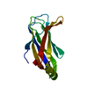
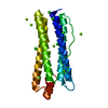
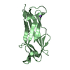
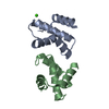
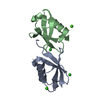
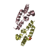
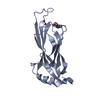
| Title | Crystal structure of Cep120 C2C domain | ||||||
 Components Components | Cep120 | ||||||
 Keywords Keywords | STRUCTURAL PROTEIN / Centriole / microtubule / Cep120 / C2 domain | ||||||
| Function / homology |  Function and homology information Function and homology informationpositive regulation of centriole elongation / positive regulation of establishment of protein localization / interkinetic nuclear migration / positive regulation of centrosome duplication / astral microtubule organization / positive regulation of cilium assembly / centrosome cycle / neurogenesis / centriole / cerebral cortex development / centrosome Similarity search - Function | ||||||
| Biological species |  homo sapiens (human) homo sapiens (human) | ||||||
| Method |  X-RAY DIFFRACTION / SYNCHROTRON / SAD / Resolution: 1.6 Å X-RAY DIFFRACTION / SYNCHROTRON / SAD / Resolution: 1.6 Å | ||||||
 Authors Authors | Sharma, A. / Gerard, S.F. / Olieric, N. / Steinmetz, M.O. | ||||||
 Citation Citation | Journal: J. Struct. Biol. / Year: 2018 Title: Cep120 promotes microtubule formation through a unique tubulin binding C2 domain. Authors: Sharma, A. / Gerard, S.F. / Olieric, N. / Steinmetz, M.O. | ||||||
| History |
|
- Structure visualization
| Structure viewer | Molecule: MolmilJmol/JSmol |
|---|
- Downloads & links
-Download
| PDBx/mmCIF format | 6flk.cif.gz | 202.1 KB | Display | PDBx/mmCIF format |
|---|---|---|---|---|
| PDB format | pdb6flk.ent.gz | 164.7 KB | Display | PDB format |
| PDBx/mmJSON format | 6flk.json.gz | Tree view | PDBx/mmJSON format | |
| Others |  Other downloads Other downloads |
-Validation report
| Arichive directory | https://data.pdbj.org/pub/pdb/validation_reports/fl/6flkftp://data.pdbj.org/pub/pdb/validation_reports/fl/6flk | HTTPS FTP |
|---|
-Related structure data

-Links
 PDBj
PDBj


- Assembly
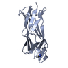
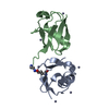
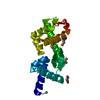
| Deposited unit | 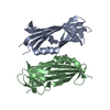
| ||||||||
|---|---|---|---|---|---|---|---|---|---|
| 1 | 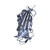
| ||||||||
| 2 | 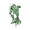
| ||||||||
| Unit cell |
|
-Components
| #1: Protein | Mass: 18537.324 Da / Num. of mol.: 2 Source method: isolated from a genetically manipulated source Source: (gene. exp.) homo sapiens (human) / Production host:  #2: Water | ChemComp-HOH / |  Mass: 18.015 Da / Num. of mol.: 384 / Source method: isolated from a natural source / Formula: H2O Mass: 18.015 Da / Num. of mol.: 384 / Source method: isolated from a natural source / Formula: H2O |
|---|
-Experimental details
-Experiment
| Experiment | Method: X-RAY DIFFRACTION / Number of used crystals: 1 |
|---|
- Sample preparation
| Crystal | Density Matthews: 2.39 Å3/Da / Density % sol: 48.55 % |
|---|---|
| Crystal grow | Temperature: 293 K / Method: vapor diffusion, sitting drop Details: 0.03M magnesium chloride hexahydrate; 0.03M calcium chloride dehydrate, 0.1M Tris(Base)/BICINE pH 8.5, 20% v/v Ethylene glycol and 10 % w/v PEG 8000 |
-Data collection
| Diffraction | Mean temperature: 100 K |
|---|---|
| Diffraction source | Source: SYNCHROTRON / Site: SLS  / Beamline: X06DA / Wavelength: 1 Å / Beamline: X06DA / Wavelength: 1 Å |
| Detector | Type: PSI PILATUS 6M / Detector: PIXEL / Date: Oct 30, 2016 |
| Radiation | Protocol: SINGLE WAVELENGTH / Monochromatic (M) / Laue (L): M / Scattering type: x-ray |
| Radiation wavelength | Wavelength: 1 Å / Relative weight: 1 |
| Reflection | Resolution: 1.6→39.97 Å / Num. obs: 42539 / % possible obs: 93.83 % / Redundancy: 1.8 % / Biso Wilson estimate: 15.125252799 Å2 / CC1/2: 0.994 / Rmerge(I) obs: 0.0819 / Rrim(I) all: 0.1158 / Net I/σ(I): 6.87 |
| Reflection shell | Resolution: 1.6→1.657 Å / Redundancy: 1.6 % / Rmerge(I) obs: 0.6926 / Mean I/σ(I) obs: 1.08 / Num. unique obs: 3744 / CC1/2: 0.439 / Rrim(I) all: 0.9794 / % possible all: 83.09 |
- Processing
| Software |
| ||||||||||||||||||||||||||||||||||||||||||||||||||||||||||||||||||||||||||||||||||||||||||||||||||||||||||||||||||||||||||||||||||||||||||||||||||||||||||||||||||||||||||||||||||||||
|---|---|---|---|---|---|---|---|---|---|---|---|---|---|---|---|---|---|---|---|---|---|---|---|---|---|---|---|---|---|---|---|---|---|---|---|---|---|---|---|---|---|---|---|---|---|---|---|---|---|---|---|---|---|---|---|---|---|---|---|---|---|---|---|---|---|---|---|---|---|---|---|---|---|---|---|---|---|---|---|---|---|---|---|---|---|---|---|---|---|---|---|---|---|---|---|---|---|---|---|---|---|---|---|---|---|---|---|---|---|---|---|---|---|---|---|---|---|---|---|---|---|---|---|---|---|---|---|---|---|---|---|---|---|---|---|---|---|---|---|---|---|---|---|---|---|---|---|---|---|---|---|---|---|---|---|---|---|---|---|---|---|---|---|---|---|---|---|---|---|---|---|---|---|---|---|---|---|---|---|---|---|---|---|
| Refinement | Method to determine structure: SAD / Resolution: 1.6→39.97 Å / SU ML: 0.24 / Cross valid method: FREE R-VALUE / σ(F): 1.2 / Phase error: 27.61
| ||||||||||||||||||||||||||||||||||||||||||||||||||||||||||||||||||||||||||||||||||||||||||||||||||||||||||||||||||||||||||||||||||||||||||||||||||||||||||||||||||||||||||||||||||||||
| Solvent computation | Shrinkage radii: 0.9 Å / VDW probe radii: 1.11 Å | ||||||||||||||||||||||||||||||||||||||||||||||||||||||||||||||||||||||||||||||||||||||||||||||||||||||||||||||||||||||||||||||||||||||||||||||||||||||||||||||||||||||||||||||||||||||
| Refinement step | Cycle: LAST / Resolution: 1.6→39.97 Å
| ||||||||||||||||||||||||||||||||||||||||||||||||||||||||||||||||||||||||||||||||||||||||||||||||||||||||||||||||||||||||||||||||||||||||||||||||||||||||||||||||||||||||||||||||||||||
| Refine LS restraints |
| ||||||||||||||||||||||||||||||||||||||||||||||||||||||||||||||||||||||||||||||||||||||||||||||||||||||||||||||||||||||||||||||||||||||||||||||||||||||||||||||||||||||||||||||||||||||
| LS refinement shell |
|
