 Movie
Movie Controller


+ Open data

- Basic information
| Entry | Database: PDB / ID: 6sie | ||||||
|---|---|---|---|---|---|---|---|
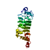
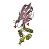
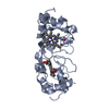
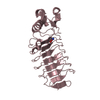
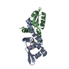
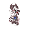
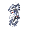
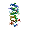
| Title | Crystal structure of the C-lobe of drosophila Arc 2 | ||||||
 Components Components | Activity-regulated cytoskeleton associated protein 2 | ||||||
 Keywords Keywords | VIRUS LIKE PARTICLE / Arc / Gag homology / atomic resolution / capsid assembly | ||||||
| Function / homology | Ty3 transposon capsid-like protein / Ty3 transposon capsid-like protein / virus-like capsid / extracellular vesicle / structural molecule activity / RNA binding / identical protein binding / Activity-regulated cytoskeleton associated protein 2 Function and homology information Function and homology information | ||||||
| Biological species |  | ||||||
| Method |  X-RAY DIFFRACTION / SYNCHROTRON / MOLECULAR REPLACEMENT / Resolution: 2.8 Å X-RAY DIFFRACTION / SYNCHROTRON / MOLECULAR REPLACEMENT / Resolution: 2.8 Å | ||||||
 Authors Authors | Hallin, E.I. / Kursula, P. | ||||||
| Funding support |  Norway, 1items Norway, 1items
| ||||||
 Citation Citation | Journal: Plos One / Year: 2021 Title: Crystal and solution structures reveal oligomerization of individual capsid homology domains of Drosophila Arc. Authors: Hallin, E.I. / Markusson, S. / Bottger, L. / Torda, A.E. / Bramham, C.R. / Kursula, P. | ||||||
| History |
|
- Structure visualization
| Structure viewer | Molecule: MolmilJmol/JSmol |
|---|
- Downloads & links
-Download
| PDBx/mmCIF format | 6sie.cif.gz | 141.7 KB | Display | PDBx/mmCIF format |
|---|---|---|---|---|
| PDB format | pdb6sie.ent.gz | 113.6 KB | Display | PDB format |
| PDBx/mmJSON format | 6sie.json.gz | Tree view | PDBx/mmJSON format | |
| Others |  Other downloads Other downloads |
-Validation report
| Arichive directory | https://data.pdbj.org/pub/pdb/validation_reports/si/6sieftp://data.pdbj.org/pub/pdb/validation_reports/si/6sie | HTTPS FTP |
|---|
-Related structure data
| Related structure data | 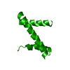 6sibC  6sidSC S: Starting model for refinement C: citing same article ( |
|---|---|
| Similar structure data |


-Links
 PDBj
PDBj- Assembly
| Deposited unit | 
| ||||||||||||
|---|---|---|---|---|---|---|---|---|---|---|---|---|---|
| 1 | 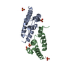
| ||||||||||||
| 2 | 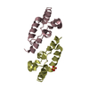
| ||||||||||||
| Unit cell |
|
-Components
| #1: Protein | Mass: 10512.886 Da / Num. of mol.: 4 Source method: isolated from a genetically manipulated source Source: (gene. exp.)  #2: Chemical | ChemComp-SO4 /   Mass: 96.063 Da / Num. of mol.: 6 / Source method: obtained synthetically / Formula: SO4 Mass: 96.063 Da / Num. of mol.: 6 / Source method: obtained synthetically / Formula: SO4#3: Water | ChemComp-HOH / |  Mass: 18.015 Da / Num. of mol.: 29 / Source method: isolated from a natural source / Formula: H2O Mass: 18.015 Da / Num. of mol.: 29 / Source method: isolated from a natural source / Formula: H2OHas ligand of interest | N | Has protein modification | Y | |
|---|
-Experimental details
-Experiment
| Experiment | Method: X-RAY DIFFRACTION / Number of used crystals: 1 |
|---|
- Sample preparation
| Crystal | Density Matthews: 4.34 Å3/Da / Density % sol: 71.63 % |
|---|---|
| Crystal grow | Temperature: 293 K / Method: vapor diffusion, sitting drop / pH: 8.5 Details: 1.25 M ammonium sulphate, 100 mM Tris pH 8.5, 200 mM lithium sulphate |
-Data collection
| Diffraction | Mean temperature: 100 K / Serial crystal experiment: N |
|---|---|
| Diffraction source | Source: SYNCHROTRON / Site: PETRA III, DESY  / Beamline: P11 / Wavelength: 1.0332 Å / Beamline: P11 / Wavelength: 1.0332 Å |
| Detector | Type: DECTRIS PILATUS 6M-F / Detector: PIXEL / Date: Jun 14, 2019 |
| Radiation | Protocol: SINGLE WAVELENGTH / Monochromatic (M) / Laue (L): M / Scattering type: x-ray |
| Radiation wavelength | Wavelength: 1.0332 Å / Relative weight: 1 |
| Reflection | Resolution: 2.8→50 Å / Num. obs: 19015 / % possible obs: 99.8 % / Redundancy: 14.3 % / Biso Wilson estimate: 49.8 Å2 / CC1/2: 0.992 / Rrim(I) all: 0.357 / Rsym value: 0.344 / Net I/σ(I): 8.7 |
| Reflection shell | Resolution: 2.8→2.87 Å / Redundancy: 13.9 % / Mean I/σ(I) obs: 0.9 / Num. unique obs: 1361 / CC1/2: 0.433 / Rrim(I) all: 3.152 / Rsym value: 3.036 / % possible all: 98.3 |
- Processing
| Software |
| ||||||||||||||||||||||||
|---|---|---|---|---|---|---|---|---|---|---|---|---|---|---|---|---|---|---|---|---|---|---|---|---|---|
| Refinement | Method to determine structure: MOLECULAR REPLACEMENT Starting model: 6SID Resolution: 2.8→50 Å / Cross valid method: FREE R-VALUE
| ||||||||||||||||||||||||
| Displacement parameters | Biso mean: 50.77 Å2 | ||||||||||||||||||||||||
| Refinement step | Cycle: LAST / Resolution: 2.8→50 Å
| ||||||||||||||||||||||||
| Refine LS restraints |
|
