- PDB-1q4o: The structure of the polo box domain of human Plk1 -
+
Open data
ID or keywords:
Loading...
-
Basic information
Entry
Database: PDB / ID: 1q4o
Title
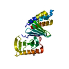
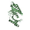
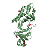
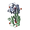
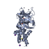
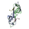
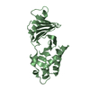
The structure of the polo box domain of human Plk1
Components
Serine/threonine-protein kinase PLK
Keywords
TRANSFERASE / Serine/threonine-protein kinase / ATP-binding / Nuclear protein
Function / homology
Function and homology information
positive regulation of mitotic nuclear envelope disassembly / Mitotic Telophase/Cytokinesis / regulation of protein localization to cell cortex / Mitotic Metaphase/Anaphase Transition / Activation of NIMA Kinases NEK9, NEK6, NEK7 / polo kinase / Phosphorylation of Emi1 / mitotic nuclear membrane disassembly / metaphase/anaphase transition of mitotic cell cycle / anaphase-promoting complex binding ...positive regulation of mitotic nuclear envelope disassembly / Mitotic Telophase/Cytokinesis / regulation of protein localization to cell cortex / Mitotic Metaphase/Anaphase Transition / Activation of NIMA Kinases NEK9, NEK6, NEK7 / polo kinase / Phosphorylation of Emi1 / mitotic nuclear membrane disassembly / metaphase/anaphase transition of mitotic cell cycle / anaphase-promoting complex binding / synaptonemal complex / Phosphorylation of the APC/C / astral microtubule organization / Golgi inheritance / mitotic cleavage furrow formation / outer kinetochore / microtubule bundle formation / mitotic chromosome condensation / double-strand break repair via alternative nonhomologous end joining / regulation of mitotic spindle assembly / Polo-like kinase mediated events / Golgi Cisternae Pericentriolar Stack Reorganization / positive regulation of mitotic metaphase/anaphase transition / positive regulation of ubiquitin-dependent protein catabolic process / centrosome cycle / regulation of mitotic metaphase/anaphase transition / sister chromatid cohesion / mitotic spindle assembly checkpoint signaling / mitotic spindle pole / regulation of mitotic cell cycle phase transition / spindle midzone / mitotic G2 DNA damage checkpoint signaling / regulation of anaphase-promoting complex-dependent catabolic process / mitotic cytokinesis / mitotic sister chromatid segregation / negative regulation of double-strand break repair via homologous recombination / Regulation of MITF-M-dependent genes involved in cell cycle and proliferation / Cyclin A/B1/B2 associated events during G2/M transition / centriolar satellite / protein localization to chromatin / Loss of Nlp from mitotic centrosomes / Loss of proteins required for interphase microtubule organization from the centrosome / Amplification of signal from unattached kinetochores via a MAD2 inhibitory signal / Recruitment of mitotic centrosome proteins and complexes / Recruitment of NuMA to mitotic centrosomes / centriole / Anchoring of the basal body to the plasma membrane / regulation of mitotic cell cycle / Mitotic Prometaphase / EML4 and NUDC in mitotic spindle formation / AURKA Activation by TPX2 / Resolution of Sister Chromatid Cohesion / mitotic spindle organization / Condensation of Prophase Chromosomes / regulation of cytokinesis / establishment of protein localization / RHO GTPases Activate Formins / positive regulation of protein localization to nucleus / APC/C:Cdh1 mediated degradation of Cdc20 and other APC/C:Cdh1 targeted proteins in late mitosis/early G1 / G2/M transition of mitotic cell cycle / kinetochore / spindle / spindle pole / The role of GTSE1 in G2/M progression after G2 checkpoint / Separation of Sister Chromatids / Regulation of PLK1 Activity at G2/M Transition / double-strand break repair / mitotic cell cycle / positive regulation of proteasomal ubiquitin-dependent protein catabolic process / microtubule cytoskeleton / midbody / microtubule binding / protein phosphorylation / protein kinase activity / regulation of cell cycle / protein serine kinase activity / protein serine/threonine kinase activity / centrosome / protein kinase binding / negative regulation of apoptotic process / chromatin / magnesium ion binding / negative regulation of transcription by RNA polymerase II / nucleoplasm / ATP binding / identical protein binding / nucleus / cytosol / cytoplasm Similarity search - Function
POLO box domain / Polo-like kinase 1, catalytic domain / Second polo-box domain / First polo-box domain / POLO box domain superfamily / POLO box duplicated region / Arylsulfatase, C-terminal domain / POLO box domain / POLO box domain profile. / Serine/threonine-protein kinase, active site ...POLO box domain / Polo-like kinase 1, catalytic domain / Second polo-box domain / First polo-box domain / POLO box domain superfamily / POLO box duplicated region / Arylsulfatase, C-terminal domain / POLO box domain / POLO box domain profile. / Serine/threonine-protein kinase, active site / Serine/Threonine protein kinases active-site signature. / Protein kinase domain / Serine/Threonine protein kinases, catalytic domain / Protein kinase, ATP binding site / Protein kinases ATP-binding region signature. / Protein kinase domain profile. / Protein kinase domain / Protein kinase-like domain superfamily / 2-Layer Sandwich / Alpha Beta Similarity search - Domain/homology
Serine/threonine-proteinkinasePLK / PLK-1 / Serine-threonine protein kinase 13 / STPK13
Mass: 27272.113 Da / Num. of mol.: 2 / Fragment: Polo box domain of Plk1 Source method: isolated from a genetically manipulated source Source: (gene. exp.) Homo sapiens (human) / Gene: PLK OR PLK1 / Plasmid: pGEX-6P1 / Production host: Escherichia coli (E. coli) / Strain (production host): B834 (DE3) pLysS References: UniProt: P53350, Transferases; Transferring phosphorus-containing groups; Phosphotransferases with an alcohol group as acceptor
Resolution: 2.2→2.26 Å / Redundancy: 2.2 % / Mean I/σ(I) obs: 3.6 / Rsym value: 0.146 / % possible all: 92
Reflection
*PLUS
Num. measured all: 103961 / Rmerge(I) obs: 0.05
Reflection shell
*PLUS
% possible obs: 92 % / Rmerge(I) obs: 0.146
-
Processing
Software
Name
Version
Classification
MOSFLM
datareduction
SCALA
datascaling
SOLVE
phasing
REFMAC
5.1
refinement
CCP4
(SCALA)
datascaling
Refinement
Method to determine structure: SAD Starting model: Model derived from a SOLVE phased map from data of a Se-Met derivative Resolution: 2.2→20 Å / Isotropic thermal model: isotropic / Cross valid method: THROUGHOUT / σ(F): 0 / σ(I): 0 / Stereochemistry target values: Engh & Huber
Rfactor
Num. reflection
% reflection
Selection details
Rfree
0.288
1041
-
Random
Rwork
0.198
-
-
-
all
0.203
20156
-
-
obs
0.198
19093
95.2 %
-
Displacement parameters
Biso mean: 31.2 Å2
Baniso -1
Baniso -2
Baniso -3
1-
-0.4 Å2
0.22 Å2
0.45 Å2
2-
-
-0.88 Å2
-1.86 Å2
3-
-
-
1.22 Å2
Refinement step
Cycle: LAST / Resolution: 2.2→20 Å
Protein
Nucleic acid
Ligand
Solvent
Total
Num. atoms
3395
0
0
227
3622
Refine LS restraints
Refine-ID
Type
Dev ideal
X-RAY DIFFRACTION
r_bond_refined_d
0.017
X-RAY DIFFRACTION
r_bond_other_d
0.002
X-RAY DIFFRACTION
r_angle_refined_deg
1.72
X-RAY DIFFRACTION
r_angle_other_deg
0.9
X-RAY DIFFRACTION
r_dihedral_angle_1_deg
7.62
LS refinement shell
Resolution: 2.2→2.257 Å
Rfactor
Num. reflection
% reflection
Rfree
0.352
78
-
Rwork
0.227
-
-
obs
-
1266
92 %
Refine LS restraints
*PLUS
Refine-ID
Type
Dev ideal
X-RAY DIFFRACTION
bond_d
0.017
X-RAY DIFFRACTION
angle_d
X-RAY DIFFRACTION
angle_deg
1.72
+
About Yorodumi
-
News
-
Feb 9, 2022. New format data for meta-information of EMDB entries
New format data for meta-information of EMDB entries
Version 3 of the EMDB header file is now the official format.
The previous official version 1.9 will be removed from the archive.
In the structure databanks used in Yorodumi, some data are registered as the other names, "COVID-19 virus" and "2019-nCoV". Here are the details of the virus and the list of structure data.
Jan 31, 2019. EMDB accession codes are about to change! (news from PDBe EMDB page)
EMDB accession codes are about to change! (news from PDBe EMDB page)
The allocation of 4 digits for EMDB accession codes will soon come to an end. Whilst these codes will remain in use, new EMDB accession codes will include an additional digit and will expand incrementally as the available range of codes is exhausted. The current 4-digit format prefixed with “EMD-” (i.e. EMD-XXXX) will advance to a 5-digit format (i.e. EMD-XXXXX), and so on. It is currently estimated that the 4-digit codes will be depleted around Spring 2019, at which point the 5-digit format will come into force.
The EM Navigator/Yorodumi systems omit the EMD- prefix.
Related info.:Q: What is EMD? / ID/Accession-code notation in Yorodumi/EM Navigator
Yorodumi is a browser for structure data from EMDB, PDB, SASBDB, etc.
This page is also the successor to EM Navigator detail page, and also detail information page/front-end page for Omokage search.
The word "yorodu" (or yorozu) is an old Japanese word meaning "ten thousand". "mi" (miru) is to see.
Related info.:EMDB / PDB / SASBDB / Comparison of 3 databanks / Yorodumi Search / Aug 31, 2016. New EM Navigator & Yorodumi / Yorodumi Papers / Jmol/JSmol / Function and homology information / Changes in new EM Navigator and Yorodumi
 Movie
Movie Controller


 Open data
 Basic information
 Components
 Keywords
 Function and homology information
 Homo sapiens (human)
 X-RAY DIFFRACTION /
  Authors
 Citation
 Structure visualization
 Downloads & links
 Other downloads


 PDBj
PDBj


 Assembly
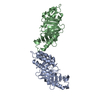


 Mass: 18.015 Da / Num. of mol.: 227 / Source method: isolated from a natural source / Formula: H2O
 Sample preparation

 Processing