 Movie
Movie Controller

[English] 日本語
 Yorodumi
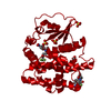
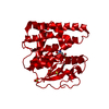
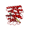
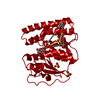
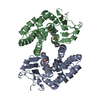
- PDB-5ueh: Structure of GSTO1 covalently conjugated to quinolinic acid fluor... -
+ Open data

- Basic information
| Entry | Database: PDB / ID: 5ueh | ||||||
|---|---|---|---|---|---|---|---|
| Title | Structure of GSTO1 covalently conjugated to quinolinic acid fluorosulfate | ||||||
 Components Components | Glutathione S-transferase omega-1 | ||||||
 Keywords Keywords | TRANSFERASE/OXIDOREDUCTASE / arylfluorosulfate / GST / covalent inhibitor / TRANSFERASE-OXIDOREDUCTASE complex | ||||||
| Function / homology |  Function and homology information Function and homology informationpositive regulation of skeletal muscle contraction by regulation of release of sequestered calcium ion / methylarsonate reductase / methylarsonate reductase activity / Vitamin C (ascorbate) metabolism / L-ascorbic acid metabolic process / glutathione dehydrogenase (ascorbate) / glutathione dehydrogenase (ascorbate) activity / cellular response to arsenic-containing substance / Methylation / Glutathione conjugation ...positive regulation of skeletal muscle contraction by regulation of release of sequestered calcium ion / methylarsonate reductase / methylarsonate reductase activity / Vitamin C (ascorbate) metabolism / L-ascorbic acid metabolic process / glutathione dehydrogenase (ascorbate) / glutathione dehydrogenase (ascorbate) activity / cellular response to arsenic-containing substance / Methylation / Glutathione conjugation / negative regulation of release of sequestered calcium ion into cytosol / glutathione transferase / glutathione transferase activity / xenobiotic catabolic process / regulation of release of sequestered calcium ion into cytosol by sarcoplasmic reticulum / regulation of cardiac muscle contraction by regulation of the release of sequestered calcium ion / Gene and protein expression by JAK-STAT signaling after Interleukin-12 stimulation / positive regulation of release of sequestered calcium ion into cytosol / glutathione metabolic process / oxidoreductase activity / extracellular exosome / cytoplasm / cytosol Similarity search - Function | ||||||
| Biological species |  Homo sapiens (human) Homo sapiens (human) | ||||||
| Method |  X-RAY DIFFRACTION / MOLECULAR REPLACEMENT / Resolution: 2 Å X-RAY DIFFRACTION / MOLECULAR REPLACEMENT / Resolution: 2 Å | ||||||
 Authors Authors | Mortenson, D.E. / Wilson, I.A. / Kelly, J.W. | ||||||
| Funding support |  United States, 1items United States, 1items
| ||||||
 Citation Citation | Journal: J. Am. Chem. Soc. / Year: 2018 Title: "Inverse Drug Discovery" Strategy To Identify Proteins That Are Targeted by Latent Electrophiles As Exemplified by Aryl Fluorosulfates. Authors: Mortenson, D.E. / Brighty, G.J. / Plate, L. / Bare, G. / Chen, W. / Li, S. / Wang, H. / Cravatt, B.F. / Forli, S. / Powers, E.T. / Sharpless, K.B. / Wilson, I.A. / Kelly, J.W. | ||||||
| History |
|
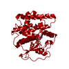
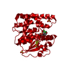
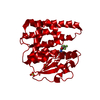
- Structure visualization
| Structure viewer | Molecule: MolmilJmol/JSmol |
|---|
- Downloads & links
-Download
| PDBx/mmCIF format | 5ueh.cif.gz | 70.2 KB | Display | PDBx/mmCIF format |
|---|---|---|---|---|
| PDB format | pdb5ueh.ent.gz | 50.5 KB | Display | PDB format |
| PDBx/mmJSON format | 5ueh.json.gz | Tree view | PDBx/mmJSON format | |
| Others |  Other downloads Other downloads |
-Validation report
| Arichive directory | https://data.pdbj.org/pub/pdb/validation_reports/ue/5uehftp://data.pdbj.org/pub/pdb/validation_reports/ue/5ueh | HTTPS FTP |
|---|
-Related structure data
| Related structure data | 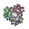 5ui4C  4is0S S: Starting model for refinement C: citing same article ( |
|---|---|
| Similar structure data |
-Links
 PDBj
PDBj


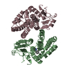
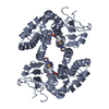
- Assembly
| Deposited unit | 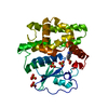
| ||||||||
|---|---|---|---|---|---|---|---|---|---|
| 1 | 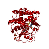
| ||||||||
| Unit cell |
|
-Components
| #1: Protein | Mass: 27686.924 Da / Num. of mol.: 1 Source method: isolated from a genetically manipulated source Source: (gene. exp.) Homo sapiens (human) / Gene: GSTO1, GSTTLP28 / Production host:  References: UniProt: P78417, glutathione transferase, glutathione dehydrogenase (ascorbate), methylarsonate reductase | ||||||
|---|---|---|---|---|---|---|---|
| #2: Chemical | ChemComp-85P / 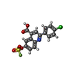  Mass: 381.763 Da / Num. of mol.: 1 / Source method: obtained synthetically / Formula: C16H9ClFNO5S Mass: 381.763 Da / Num. of mol.: 1 / Source method: obtained synthetically / Formula: C16H9ClFNO5S | ||||||
| #3: Chemical | ChemComp-SO4 /   Mass: 96.063 Da / Num. of mol.: 5 / Source method: obtained synthetically / Formula: SO4 Mass: 96.063 Da / Num. of mol.: 5 / Source method: obtained synthetically / Formula: SO4#4: Chemical | ChemComp-GOL / |   Mass: 92.094 Da / Num. of mol.: 1 / Source method: obtained synthetically / Formula: C3H8O3 Mass: 92.094 Da / Num. of mol.: 1 / Source method: obtained synthetically / Formula: C3H8O3#5: Water | ChemComp-HOH / |  Mass: 18.015 Da / Num. of mol.: 216 / Source method: isolated from a natural source / Formula: H2O Mass: 18.015 Da / Num. of mol.: 216 / Source method: isolated from a natural source / Formula: H2OHas protein modification | Y | |
-Experimental details
-Experiment
| Experiment | Method: X-RAY DIFFRACTION / Number of used crystals: 1 |
|---|
- Sample preparation
| Crystal | Density Matthews: 2.39 Å3/Da / Density % sol: 48.51 % |
|---|---|
| Crystal grow | Temperature: 298 K / Method: vapor diffusion, sitting drop / pH: 5 Details: 5 mg/mL solution of protein crystallized from precipitant solution containing 2.4 M ammonium sulfate and 0.1 M MES solution pH 5. Crystals cryoprotected by brief immersion in solution ...Details: 5 mg/mL solution of protein crystallized from precipitant solution containing 2.4 M ammonium sulfate and 0.1 M MES solution pH 5. Crystals cryoprotected by brief immersion in solution containing 1.5 M ammonium sulfate, 25% (v/v) glycerol |
-Data collection
| Diffraction | Mean temperature: 100 K |
|---|---|
| Diffraction source | Source: ROTATING ANODE / Type: RIGAKU MICROMAX-007 HF / Wavelength: 1.5418 Å |
| Detector | Type: MAR scanner 345 mm plate / Detector: IMAGE PLATE / Date: May 9, 2016 / Details: Osmic VariMax |
| Radiation | Protocol: SINGLE WAVELENGTH / Monochromatic (M) / Laue (L): M / Scattering type: x-ray |
| Radiation wavelength | Wavelength: 1.5418 Å / Relative weight: 1 |
| Reflection | Resolution: 2→49.5 Å / Num. obs: 18695 / % possible obs: 100 % / Redundancy: 8.6 % / Biso Wilson estimate: 25.6 Å2 / CC1/2: 0.998 / Rpim(I) all: 0.043 / Rsym value: 0.118 / Net I/σ(I): 11.9 |
| Reflection shell | Resolution: 2→2.11 Å / Redundancy: 8.7 % / Mean I/σ(I) obs: 2.4 / Num. unique obs: 2695 / CC1/2: 0.704 / Rpim(I) all: 0.329 / Rsym value: 0.914 / % possible all: 100 |
- Processing
| Software |
| ||||||||||||||||||||||||||||||||||||||||||||||||||||||||||||||||||||||||||||||||||||||||||||||||||||||||||||||||||||||||||||||||||||||||||||||||||||||||||||||||||||||||||||||||||||||
|---|---|---|---|---|---|---|---|---|---|---|---|---|---|---|---|---|---|---|---|---|---|---|---|---|---|---|---|---|---|---|---|---|---|---|---|---|---|---|---|---|---|---|---|---|---|---|---|---|---|---|---|---|---|---|---|---|---|---|---|---|---|---|---|---|---|---|---|---|---|---|---|---|---|---|---|---|---|---|---|---|---|---|---|---|---|---|---|---|---|---|---|---|---|---|---|---|---|---|---|---|---|---|---|---|---|---|---|---|---|---|---|---|---|---|---|---|---|---|---|---|---|---|---|---|---|---|---|---|---|---|---|---|---|---|---|---|---|---|---|---|---|---|---|---|---|---|---|---|---|---|---|---|---|---|---|---|---|---|---|---|---|---|---|---|---|---|---|---|---|---|---|---|---|---|---|---|---|---|---|---|---|---|---|
| Refinement | Method to determine structure: MOLECULAR REPLACEMENT Starting model: PDB 4IS0 Resolution: 2→49.5 Å / Cor.coef. Fo:Fc: 0.964 / Cor.coef. Fo:Fc free: 0.929 / SU B: 4.214 / SU ML: 0.114 / Cross valid method: THROUGHOUT / ESU R: 0.173 / ESU R Free: 0.165 / Stereochemistry target values: MAXIMUM LIKELIHOOD / Details: HYDROGENS HAVE BEEN ADDED IN THE RIDING POSITIONS
| ||||||||||||||||||||||||||||||||||||||||||||||||||||||||||||||||||||||||||||||||||||||||||||||||||||||||||||||||||||||||||||||||||||||||||||||||||||||||||||||||||||||||||||||||||||||
| Solvent computation | Ion probe radii: 0.8 Å / Shrinkage radii: 0.8 Å / VDW probe radii: 1.2 Å / Solvent model: MASK | ||||||||||||||||||||||||||||||||||||||||||||||||||||||||||||||||||||||||||||||||||||||||||||||||||||||||||||||||||||||||||||||||||||||||||||||||||||||||||||||||||||||||||||||||||||||
| Displacement parameters | Biso mean: 33.638 Å2
| ||||||||||||||||||||||||||||||||||||||||||||||||||||||||||||||||||||||||||||||||||||||||||||||||||||||||||||||||||||||||||||||||||||||||||||||||||||||||||||||||||||||||||||||||||||||
| Refinement step | Cycle: 1 / Resolution: 2→49.5 Å
| ||||||||||||||||||||||||||||||||||||||||||||||||||||||||||||||||||||||||||||||||||||||||||||||||||||||||||||||||||||||||||||||||||||||||||||||||||||||||||||||||||||||||||||||||||||||
| Refine LS restraints |
|