positive regulation of skeletal muscle contraction by regulation of release of sequestered calcium ion / methylarsonate reductase / methylarsonate reductase activity / Vitamin C (ascorbate) metabolism / L-ascorbic acid metabolic process / glutathione dehydrogenase (ascorbate) / glutathione dehydrogenase (ascorbate) activity / cellular response to arsenic-containing substance / Methylation / Glutathione conjugation ...positive regulation of skeletal muscle contraction by regulation of release of sequestered calcium ion / methylarsonate reductase / methylarsonate reductase activity / Vitamin C (ascorbate) metabolism / L-ascorbic acid metabolic process / glutathione dehydrogenase (ascorbate) / glutathione dehydrogenase (ascorbate) activity / cellular response to arsenic-containing substance / Methylation / Glutathione conjugation / negative regulation of release of sequestered calcium ion into cytosol / glutathione transferase / glutathione transferase activity / xenobiotic catabolic process / regulation of release of sequestered calcium ion into cytosol by sarcoplasmic reticulum / regulation of cardiac muscle contraction by regulation of the release of sequestered calcium ion / Gene and protein expression by JAK-STAT signaling after Interleukin-12 stimulation / positive regulation of release of sequestered calcium ion into cytosol / glutathione metabolic process / oxidoreductase activity / extracellular exosome / cytoplasm / cytosol Similarity search - Function
Mass: 18.015 Da / Num. of mol.: 405 / Source method: isolated from a natural source / Formula: H2O
Has protein modification
Y
-
Experimental details
-
Experiment
Experiment
Method: X-RAY DIFFRACTION / Number of used crystals: 1
-
Sample preparation
Crystal
Density Matthews: 2.38 Å3/Da / Density % sol: 48.28 %
Crystal grow
Temperature: 293 K / Method: vapor diffusion, sitting drop / pH: 6.5 Details: Complex was concentrated to 26.2 mg/mL. Crystals of the complex grew from sitting drops containing equal volumes of protein complex and well solution (22.5% PEG 3350, 90 mM MES pH 6.5 and 10 mM BaCl2).
In the structure databanks used in Yorodumi, some data are registered as the other names, "COVID-19 virus" and "2019-nCoV". Here are the details of the virus and the list of structure data.
Jan 31, 2019. EMDB accession codes are about to change! (news from PDBe EMDB page)
EMDB accession codes are about to change! (news from PDBe EMDB page)
The allocation of 4 digits for EMDB accession codes will soon come to an end. Whilst these codes will remain in use, new EMDB accession codes will include an additional digit and will expand incrementally as the available range of codes is exhausted. The current 4-digit format prefixed with “EMD-” (i.e. EMD-XXXX) will advance to a 5-digit format (i.e. EMD-XXXXX), and so on. It is currently estimated that the 4-digit codes will be depleted around Spring 2019, at which point the 5-digit format will come into force.
The EM Navigator/Yorodumi systems omit the EMD- prefix.
Related info.:Q: What is EMD? / ID/Accession-code notation in Yorodumi/EM Navigator
Yorodumi is a browser for structure data from EMDB, PDB, SASBDB, etc.
This page is also the successor to EM Navigator detail page, and also detail information page/front-end page for Omokage search.
The word "yorodu" (or yorozu) is an old Japanese word meaning "ten thousand". "mi" (miru) is to see.
Related info.:EMDB / PDB / SASBDB / Comparison of 3 databanks / Yorodumi Search / Aug 31, 2016. New EM Navigator & Yorodumi / Yorodumi Papers / Jmol/JSmol / Function and homology information / Changes in new EM Navigator and Yorodumi
 Movie
Movie Controller

 Yorodumi
 Open data
 Basic information
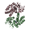
 Components
 Keywords
 Function and homology information
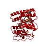
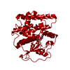
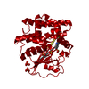
 Homo sapiens (human)
 X-RAY DIFFRACTION /
  Authors
 Citation
 Structure visualization
 Downloads & links
 Other downloads
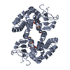


 PDBj
PDBj


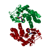
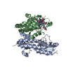
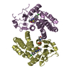
 Assembly
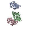


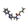
 Mass: 289.396 Da / Num. of mol.: 3 / Source method: obtained synthetically / Formula: C15H19N3OS
 Mass: 18.015 Da / Num. of mol.: 405 / Source method: isolated from a natural source / Formula: H2O
 Sample preparation
 / Beamline: 21-ID-D / Wavelength: 0.9786 Å
 Processing