- PDB-3vln: Human Glutathione Transferase O1-1 C32S Mutant in Complex with As... -
+
Open data
ID or keywords:
Loading...
-
Basic information
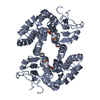
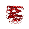
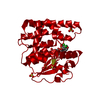
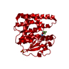
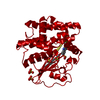
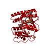
Entry
Database: PDB / ID: 3vln
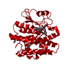
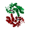
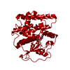
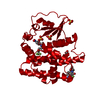
Title
Human Glutathione Transferase O1-1 C32S Mutant in Complex with Ascorbic Acid
Components
Glutathione S-transferase omega-1
Keywords
TRANSFERASE / GST fold / reductase / glutathione
Function / homology
Function and homology information
positive regulation of skeletal muscle contraction by regulation of release of sequestered calcium ion / methylarsonate reductase / methylarsonate reductase activity / Vitamin C (ascorbate) metabolism / L-ascorbic acid metabolic process / glutathione dehydrogenase (ascorbate) / glutathione dehydrogenase (ascorbate) activity / cellular response to arsenic-containing substance / Methylation / Glutathione conjugation ...positive regulation of skeletal muscle contraction by regulation of release of sequestered calcium ion / methylarsonate reductase / methylarsonate reductase activity / Vitamin C (ascorbate) metabolism / L-ascorbic acid metabolic process / glutathione dehydrogenase (ascorbate) / glutathione dehydrogenase (ascorbate) activity / cellular response to arsenic-containing substance / Methylation / Glutathione conjugation / negative regulation of release of sequestered calcium ion into cytosol / glutathione transferase / glutathione transferase activity / xenobiotic catabolic process / regulation of release of sequestered calcium ion into cytosol by sarcoplasmic reticulum / regulation of cardiac muscle contraction by regulation of the release of sequestered calcium ion / Gene and protein expression by JAK-STAT signaling after Interleukin-12 stimulation / positive regulation of release of sequestered calcium ion into cytosol / glutathione metabolic process / oxidoreductase activity / extracellular exosome / cytoplasm / cytosol Similarity search - Function
Resolution: 1.7→30 Å / Cor.coef. Fo:Fc: 0.963 / Cor.coef. Fo:Fc free: 0.952 / SU B: 4.773 / SU ML: 0.072 / Cross valid method: THROUGHOUT / ESU R: 0.106 / ESU R Free: 0.106 / Stereochemistry target values: MAXIMUM LIKELIHOOD / Details: HYDROGENS HAVE BEEN ADDED IN THE RIDING POSITIONS
Rfactor
Num. reflection
% reflection
Selection details
Rfree
0.21749
1507
5.1 %
RANDOM
Rwork
0.18043
-
-
-
obs
0.18222
28239
99.24 %
-
Solvent computation
Ion probe radii: 0.8 Å / Shrinkage radii: 0.8 Å / VDW probe radii: 1.4 Å / Solvent model: BABINET MODEL WITH MASK
Displacement parameters
Biso mean: 31.695 Å2
Baniso -1
Baniso -2
Baniso -3
1-
0.84 Å2
0.42 Å2
-0 Å2
2-
-
0.84 Å2
-0 Å2
3-
-
-
-1.25 Å2
Refinement step
Cycle: LAST / Resolution: 1.7→30 Å
Protein
Nucleic acid
Ligand
Solvent
Total
Num. atoms
1923
0
47
205
2175
Refine LS restraints
Refine-ID
Type
Dev ideal
Dev ideal target
Number
X-RAY DIFFRACTION
r_bond_refined_d
0.02
0.022
2065
X-RAY DIFFRACTION
r_bond_other_d
0.001
0.02
1465
X-RAY DIFFRACTION
r_angle_refined_deg
1.86
2.005
2797
X-RAY DIFFRACTION
r_angle_other_deg
1.02
3
3592
X-RAY DIFFRACTION
r_dihedral_angle_1_deg
5.808
5
254
X-RAY DIFFRACTION
r_dihedral_angle_2_deg
34.095
24.835
91
X-RAY DIFFRACTION
r_dihedral_angle_3_deg
15.153
15
378
X-RAY DIFFRACTION
r_dihedral_angle_4_deg
18.39
15
9
X-RAY DIFFRACTION
r_chiral_restr
0.111
0.2
293
X-RAY DIFFRACTION
r_gen_planes_refined
0.009
0.021
2247
X-RAY DIFFRACTION
r_gen_planes_other
0.001
0.02
397
X-RAY DIFFRACTION
r_nbd_refined
X-RAY DIFFRACTION
r_nbd_other
X-RAY DIFFRACTION
r_nbtor_refined
X-RAY DIFFRACTION
r_nbtor_other
X-RAY DIFFRACTION
r_xyhbond_nbd_refined
X-RAY DIFFRACTION
r_xyhbond_nbd_other
X-RAY DIFFRACTION
r_metal_ion_refined
X-RAY DIFFRACTION
r_metal_ion_other
X-RAY DIFFRACTION
r_symmetry_vdw_refined
X-RAY DIFFRACTION
r_symmetry_vdw_other
X-RAY DIFFRACTION
r_symmetry_hbond_refined
X-RAY DIFFRACTION
r_symmetry_hbond_other
X-RAY DIFFRACTION
r_symmetry_metal_ion_refined
X-RAY DIFFRACTION
r_symmetry_metal_ion_other
X-RAY DIFFRACTION
r_mcbond_it
0.993
1.5
1226
X-RAY DIFFRACTION
r_mcbond_other
0.306
1.5
485
X-RAY DIFFRACTION
r_mcangle_it
1.701
2
1988
X-RAY DIFFRACTION
r_scbond_it
2.673
3
839
X-RAY DIFFRACTION
r_scangle_it
4.149
4.5
802
X-RAY DIFFRACTION
r_rigid_bond_restr
X-RAY DIFFRACTION
r_sphericity_free
X-RAY DIFFRACTION
r_sphericity_bonded
LS refinement shell
Resolution: 1.698→1.742 Å / Total num. of bins used: 20
Rfactor
Num. reflection
% reflection
Rfree
0.257
107
-
Rwork
0.234
2020
-
obs
-
-
97.61 %
Refinement TLS params.
Method: refined / Origin x: 22.7624 Å / Origin y: -10.6049 Å / Origin z: 19.8133 Å
11
12
13
21
22
23
31
32
33
T
0.0374 Å2
0.0008 Å2
0.0009 Å2
-
0.0298 Å2
-0.0051 Å2
-
-
0.0971 Å2
L
1.0462 °2
0.225 °2
0.1619 °2
-
1.7715 °2
-0.1176 °2
-
-
3.6391 °2
S
0.0029 Å °
-0.0807 Å °
0.1159 Å °
0.09 Å °
-0.0344 Å °
0.057 Å °
-0.3497 Å °
-0.0532 Å °
0.0314 Å °
+
About Yorodumi
-
News
-
Feb 9, 2022. New format data for meta-information of EMDB entries
New format data for meta-information of EMDB entries
Version 3 of the EMDB header file is now the official format.
The previous official version 1.9 will be removed from the archive.
In the structure databanks used in Yorodumi, some data are registered as the other names, "COVID-19 virus" and "2019-nCoV". Here are the details of the virus and the list of structure data.
Jan 31, 2019. EMDB accession codes are about to change! (news from PDBe EMDB page)
EMDB accession codes are about to change! (news from PDBe EMDB page)
The allocation of 4 digits for EMDB accession codes will soon come to an end. Whilst these codes will remain in use, new EMDB accession codes will include an additional digit and will expand incrementally as the available range of codes is exhausted. The current 4-digit format prefixed with “EMD-” (i.e. EMD-XXXX) will advance to a 5-digit format (i.e. EMD-XXXXX), and so on. It is currently estimated that the 4-digit codes will be depleted around Spring 2019, at which point the 5-digit format will come into force.
The EM Navigator/Yorodumi systems omit the EMD- prefix.
Related info.:Q: What is EMD? / ID/Accession-code notation in Yorodumi/EM Navigator
Yorodumi is a browser for structure data from EMDB, PDB, SASBDB, etc.
This page is also the successor to EM Navigator detail page, and also detail information page/front-end page for Omokage search.
The word "yorodu" (or yorozu) is an old Japanese word meaning "ten thousand". "mi" (miru) is to see.
Related info.:EMDB / PDB / SASBDB / Comparison of 3 databanks / Yorodumi Search / Aug 31, 2016. New EM Navigator & Yorodumi / Yorodumi Papers / Jmol/JSmol / Function and homology information / Changes in new EM Navigator and Yorodumi
 Movie
Movie Controller

 Yorodumi
 Open data
 Basic information
 Components
 Keywords
 Function and homology information
 Homo sapiens (human)
 X-RAY DIFFRACTION /
  Authors
 Citation
 Structure visualization
 Downloads & links
 Other downloads


 PDBj
PDBj


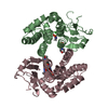
 Assembly
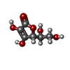

 Type: L-saccharide / Mass: 176.124 Da / Num. of mol.: 1 / Source method: obtained synthetically / Formula: C6H8O6


 Mass: 96.063 Da / Num. of mol.: 3 / Source method: obtained synthetically / Formula: SO4
 Mass: 92.094 Da / Num. of mol.: 2 / Source method: obtained synthetically / Formula: C3H8O3
 Mass: 62.068 Da / Num. of mol.: 1 / Source method: obtained synthetically / Formula: C2H6O2
 Mass: 59.044 Da / Num. of mol.: 1 / Source method: obtained synthetically / Formula: C2H3O2
 Sample preparation
 / Beamline: MX2 / Wavelength: 1 Å
 Processing