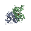
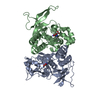
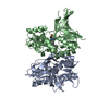
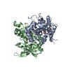
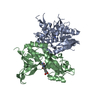
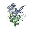
Entry Database : PDB / ID : 5u8cTitle CRYSTAL STRUCTURE OF GLUN1/GLUN2A LIGAND-BINDING DOMAIN IN COMPLEX WITH GLYCINE AND NVP-AAM077 GLUTAMATE RECEPTOR IONOTROPIC, NMDA 2A Glutamate receptor ionotropic, NMDA 1 Keywords / / Function / homology Function Domain/homology Component
/ / / / / / / / / / / / / / / / / / / / / / / / / / / / / / / / / / / / / / / / / / / / / / / / / / / / / / / / / / / / / / / / / / / / / / / / / / / / / / / / / / / / / / / / / / / / / / / / / / / / / / / / / / / / / / / / / / / / / / / / / / / / / / / / / / / / / / / Biological species Rattus norvegicus (Norway rat)Method / / / / Resolution : 1.598 Å Authors Romero-Hernandez, A. / Furukawa, H. Funding support Organization Grant number Country National Institutes of Health/National Institute of Mental Health (NIH/NIMH) MH085926 National Institutes of Health/National Institute of General Medical Sciences (NIH/NIGMS) GM105730
Journal : Mol. Pharmacol. / Year : 2017Title : Novel Mode of Antagonist Binding in NMDA Receptors Revealed by the Crystal Structure of the GluN1-GluN2A Ligand-Binding Domain Complexed to NVP-AAM077.Authors : Romero-Hernandez, A. / Furukawa, H. History Deposition Dec 14, 2016 Deposition site / Processing site Revision 1.0 May 17, 2017 Provider / Type Revision 1.1 Jun 7, 2017 Group / OtherRevision 1.2 Sep 27, 2017 Group / Category / Item Revision 1.3 Nov 27, 2019 Group / Category / Item Revision 1.4 Oct 4, 2023 Group / Database references / Refinement descriptionCategory chem_comp_atom / chem_comp_bond ... chem_comp_atom / chem_comp_bond / database_2 / pdbx_initial_refinement_model Item / _database_2.pdbx_database_accessionRevision 1.5 Oct 30, 2024 Group / Category / pdbx_modification_feature
Show all Show less
 Movie
Movie Controller

 Yorodumi
 Open data
 Basic information
 Components
 Keywords
 Function and homology information
 X-RAY DIFFRACTION /
  Authors
 United States, 2items
  Citation
 Structure visualization
 Downloads & links
 Other downloads
 PDBj
PDBj


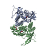
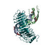
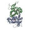
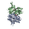
 Assembly
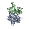


 Type: peptide linking / Mass: 75.067 Da / Num. of mol.: 1 / Source method: obtained synthetically / Formula: C2H5NO2
 Mass: 92.094 Da / Num. of mol.: 1 / Source method: obtained synthetically / Formula: C3H8O3
 Mass: 454.212 Da / Num. of mol.: 1 / Source method: obtained synthetically / Formula: C17H17BrN3O5P
 Sample preparation
 Processing