 Movie
Movie Controller

[English] 日本語
 Yorodumi
- PDB-6usu: Crystal structure of GluN1/GluN2A ligand-binding domain in comple... -
+ Open data

- Basic information
| Entry | Database: PDB / ID: 6usu | |||||||||
|---|---|---|---|---|---|---|---|---|---|---|
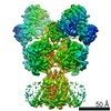
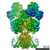
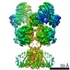
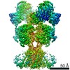
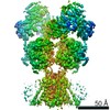
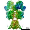
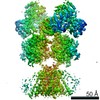
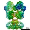
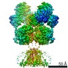
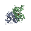
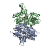
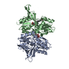
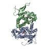
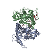
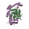
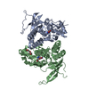
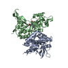
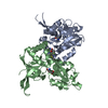
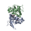
| Title | Crystal structure of GluN1/GluN2A ligand-binding domain in complex with L689,560 and glutamate | |||||||||
 Components Components |
| |||||||||
 Keywords Keywords | METAL TRANSPORT / NMDARs / LBD / Ion channels | |||||||||
| Function / homology |  Function and homology information Function and homology informationregulation of response to alcohol / response to ammonium ion / neurotransmitter receptor transport, plasma membrane to endosome / receptor recycling / response to environmental enrichment / directional locomotion / pons maturation / EPHB-mediated forward signaling / Assembly and cell surface presentation of NMDA receptors / positive regulation of Schwann cell migration ...regulation of response to alcohol / response to ammonium ion / neurotransmitter receptor transport, plasma membrane to endosome / receptor recycling / response to environmental enrichment / directional locomotion / pons maturation / EPHB-mediated forward signaling / Assembly and cell surface presentation of NMDA receptors / positive regulation of Schwann cell migration / regulation of cell communication / auditory behavior / cellular response to magnesium ion / olfactory learning / response to other organism / serotonin metabolic process / response to hydrogen sulfide / dendritic branch / conditioned taste aversion / response to methylmercury / protein localization to postsynaptic membrane / regulation of ARF protein signal transduction / suckling behavior / transmitter-gated monoatomic ion channel activity / response to manganese ion / sleep / response to carbohydrate / regulation of respiratory gaseous exchange / cellular response to lipid / propylene metabolic process / response to glycine / dendritic spine organization / locomotion / cellular response to dsRNA / regulation of NMDA receptor activity / RAF/MAP kinase cascade / positive regulation of inhibitory postsynaptic potential / neurotransmitter receptor complex / response to amine / Synaptic adhesion-like molecules / response to glycoside / regulation of monoatomic cation transmembrane transport / NMDA glutamate receptor activity / NMDA selective glutamate receptor complex / glutamate binding / voltage-gated monoatomic cation channel activity / ligand-gated sodium channel activity / glutamate receptor signaling pathway / neuromuscular process / regulation of axonogenesis / calcium ion transmembrane import into cytosol / regulation of dendrite morphogenesis / male mating behavior / protein heterotetramerization / regulation of synapse assembly / response to morphine / spinal cord development / glycine binding / dopamine metabolic process / startle response / cellular response to zinc ion / positive regulation of reactive oxygen species biosynthetic process / response to lithium ion / parallel fiber to Purkinje cell synapse / monoatomic ion channel complex / regulation of postsynaptic membrane potential / positive regulation of calcium ion transport into cytosol / associative learning / cellular response to glycine / modulation of excitatory postsynaptic potential / positive regulation of dendritic spine maintenance / monoatomic cation transmembrane transport / action potential / response to light stimulus / Unblocking of NMDA receptors, glutamate binding and activation / positive regulation of protein targeting to membrane / regulation of neuronal synaptic plasticity / monoatomic cation transport / glutamate receptor binding / social behavior / ligand-gated monoatomic ion channel activity / multicellular organismal response to stress / neuron development / conditioned place preference / prepulse inhibition / phosphatase binding / long-term memory / postsynaptic density, intracellular component / response to fungicide / synaptic cleft / monoatomic cation channel activity / calcium ion homeostasis / cellular response to manganese ion / glutamate-gated receptor activity / positive regulation of synaptic transmission, glutamatergic / glutamate-gated calcium ion channel activity / presynaptic active zone membrane / cell adhesion molecule binding / neurogenesis / excitatory synapse Similarity search - Function | |||||||||
| Biological species |  | |||||||||
| Method |  X-RAY DIFFRACTION / SYNCHROTRON / MOLECULAR REPLACEMENT / Resolution: 2.092 Å X-RAY DIFFRACTION / SYNCHROTRON / MOLECULAR REPLACEMENT / Resolution: 2.092 Å | |||||||||
 Authors Authors | Romero-Hernandez, A. / Tajima, N. / Chou, T. / Furukawa, H. | |||||||||
| Funding support |  United States, 2items United States, 2items
| |||||||||
 Citation Citation | Journal: Cell / Year: 2020 Title: Structural Basis of Functional Transitions in Mammalian NMDA Receptors. Authors: Tsung-Han Chou / Nami Tajima / Annabel Romero-Hernandez / Hiro Furukawa / Abstract: Excitatory neurotransmission meditated by glutamate receptors including N-methyl-D-aspartate receptors (NMDARs) is pivotal to brain development and function. NMDARs are heterotetramers composed of ...Excitatory neurotransmission meditated by glutamate receptors including N-methyl-D-aspartate receptors (NMDARs) is pivotal to brain development and function. NMDARs are heterotetramers composed of GluN1 and GluN2 subunits, which bind glycine and glutamate, respectively, to activate their ion channels. Despite importance in brain physiology, the precise mechanisms by which activation and inhibition occur via subunit-specific binding of agonists and antagonists remain largely unknown. Here, we show the detailed patterns of conformational changes and inter-subunit and -domain reorientation leading to agonist-gating and subunit-dependent competitive inhibition by providing multiple structures in distinct ligand states at 4 Å or better. The structures reveal that activation and competitive inhibition by both GluN1 and GluN2 antagonists occur by controlling the tension of the linker between the ligand-binding domain and the transmembrane ion channel of the GluN2 subunit. Our results provide detailed mechanistic insights into NMDAR pharmacology, activation, and inhibition, which are fundamental to the brain physiology. | |||||||||
| History |
|
- Structure visualization
| Structure viewer | Molecule: MolmilJmol/JSmol |
|---|
- Downloads & links
-Download
| PDBx/mmCIF format | 6usu.cif.gz | 131.9 KB | Display | PDBx/mmCIF format |
|---|---|---|---|---|
| PDB format | pdb6usu.ent.gz | 98.5 KB | Display | PDB format |
| PDBx/mmJSON format | 6usu.json.gz | Tree view | PDBx/mmJSON format | |
| Others |  Other downloads Other downloads |
-Validation report
| Arichive directory | https://data.pdbj.org/pub/pdb/validation_reports/us/6usuftp://data.pdbj.org/pub/pdb/validation_reports/us/6usu | HTTPS FTP |
|---|
-Related structure data
| Related structure data | 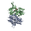 6usvC  6whrC 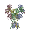 6whsC 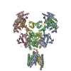 6whtC 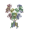 6whuC  6whvC  6whwC  6whxC  6whyC  6wi0C 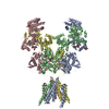 6wi1C  4nf8S S: Starting model for refinement C: citing same article ( |
|---|---|
| Similar structure data |

-Links
 PDBj
PDBj


- Assembly
| Deposited unit | 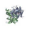
| ||||||||
|---|---|---|---|---|---|---|---|---|---|
| 1 |
| ||||||||
| Unit cell |
|
-Components
| #1: Protein | Mass: 33340.031 Da / Num. of mol.: 1 / Fragment: UNP residues 415-565, 684-821 Source method: isolated from a genetically manipulated source Source: (gene. exp.)  |
|---|---|
| #2: Protein | Mass: 31785.299 Da / Num. of mol.: 1 / Fragment: UNP residues 402-539, 661-802 Source method: isolated from a genetically manipulated source Source: (gene. exp.) |
| #3: Chemical | ChemComp-QGM / (  Mass: 380.225 Da / Num. of mol.: 1 / Source method: obtained synthetically / Formula: C17H15Cl2N3O3 / Feature type: SUBJECT OF INVESTIGATION Mass: 380.225 Da / Num. of mol.: 1 / Source method: obtained synthetically / Formula: C17H15Cl2N3O3 / Feature type: SUBJECT OF INVESTIGATION |
| #4: Chemical | ChemComp-GLU /   Type: L-peptide linking / Mass: 147.129 Da / Num. of mol.: 1 / Source method: obtained synthetically / Formula: C5H9NO4 / Feature type: SUBJECT OF INVESTIGATION Type: L-peptide linking / Mass: 147.129 Da / Num. of mol.: 1 / Source method: obtained synthetically / Formula: C5H9NO4 / Feature type: SUBJECT OF INVESTIGATION |
| #5: Water | ChemComp-HOH /  Mass: 18.015 Da / Num. of mol.: 249 / Source method: isolated from a natural source / Formula: H2O Mass: 18.015 Da / Num. of mol.: 249 / Source method: isolated from a natural source / Formula: H2O |
| Has ligand of interest | Y |
| Has protein modification | Y |
-Experimental details
-Experiment
| Experiment | Method: X-RAY DIFFRACTION / Number of used crystals: 1 |
|---|
- Sample preparation
| Crystal | Density Matthews: 2.48 Å3/Da / Density % sol: 50.44 % |
|---|---|
| Crystal grow | Temperature: 291 K / Method: evaporation / pH: 7 Details: 0.2 M HEPES, pH 7.0, 60-90 mM sodium chloride, 15-20% PEG2000 MME |
-Data collection
| Diffraction | Mean temperature: 100 K / Serial crystal experiment: N |
|---|---|
| Diffraction source | Source: SYNCHROTRON / Site: APS / Beamline: 23-ID-B / Wavelength: 1.1 Å |
| Detector | Type: DECTRIS EIGER X 16M / Detector: PIXEL / Date: Apr 6, 2013 |
| Radiation | Monochromator: Double crystal cryo-cooled Si(111) / Protocol: SINGLE WAVELENGTH / Monochromatic (M) / Laue (L): M / Scattering type: x-ray |
| Radiation wavelength | Wavelength: 1.1 Å / Relative weight: 1 |
| Reflection | Resolution: 2.09→40 Å / Num. obs: 38873 / % possible obs: 99.9 % / Redundancy: 7.6 % / Biso Wilson estimate: 25.56 Å2 / Rmerge(I) obs: 0.117 / Net I/σ(I): 17.1 |
| Reflection shell | Resolution: 2.09→2.18 Å / Redundancy: 6 % / Rmerge(I) obs: 0.62 / Num. unique obs: 3781 / CC1/2: 0.814 / % possible all: 99.8 |
- Processing
| Software |
| ||||||||||||||||||||||||||||||||||||||||||||||||||||||||||||||||||||||||||||||||||||
|---|---|---|---|---|---|---|---|---|---|---|---|---|---|---|---|---|---|---|---|---|---|---|---|---|---|---|---|---|---|---|---|---|---|---|---|---|---|---|---|---|---|---|---|---|---|---|---|---|---|---|---|---|---|---|---|---|---|---|---|---|---|---|---|---|---|---|---|---|---|---|---|---|---|---|---|---|---|---|---|---|---|---|---|---|---|
| Refinement | Method to determine structure: MOLECULAR REPLACEMENT Starting model: PDB entry 4NF8 Resolution: 2.092→38.161 Å / SU ML: 0.23 / Cross valid method: THROUGHOUT / σ(F): 1.36 / Phase error: 21.77
| ||||||||||||||||||||||||||||||||||||||||||||||||||||||||||||||||||||||||||||||||||||
| Solvent computation | Shrinkage radii: 0.9 Å / VDW probe radii: 1.11 Å | ||||||||||||||||||||||||||||||||||||||||||||||||||||||||||||||||||||||||||||||||||||
| Displacement parameters | Biso max: 77.3 Å2 / Biso mean: 27.2215 Å2 / Biso min: 12.93 Å2 | ||||||||||||||||||||||||||||||||||||||||||||||||||||||||||||||||||||||||||||||||||||
| Refinement step | Cycle: final / Resolution: 2.092→38.161 Å
| ||||||||||||||||||||||||||||||||||||||||||||||||||||||||||||||||||||||||||||||||||||
| LS refinement shell | Refine-ID: X-RAY DIFFRACTION / Rfactor Rfree error: 0
|
