 Movie
Movie Controller

[English] 日本語
 Yorodumi
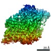
- PDB-2es4: Crystal structure of the Burkholderia glumae lipase-specific fold... -
+ Open data

- Basic information
| Entry | Database: PDB / ID: 2es4 | ||||||
|---|---|---|---|---|---|---|---|
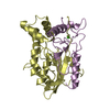
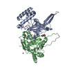
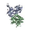
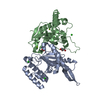
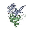
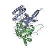
| Title | Crystal structure of the Burkholderia glumae lipase-specific foldase in complex with its cognate lipase | ||||||
 Components Components |
| ||||||
 Keywords Keywords | HYDROLASE / protein-protein complex / steric chaperone / triacylglycerol hydrolase / all alpha helix protein / a/b hydrolase fold / extensive interaction area | ||||||
| Function / homology |  Function and homology information Function and homology informationtriacylglycerol lipase / triacylglycerol lipase activity / lipid catabolic process / : / protein folding / extracellular region / metal ion binding / plasma membrane Similarity search - Function | ||||||
| Biological species |  Burkholderia glumae (bacteria) Burkholderia glumae (bacteria) | ||||||
| Method |  X-RAY DIFFRACTION / SYNCHROTRON / MOLECULAR REPLACEMENT / Resolution: 1.85 Å X-RAY DIFFRACTION / SYNCHROTRON / MOLECULAR REPLACEMENT / Resolution: 1.85 Å | ||||||
 Authors Authors | Pauwels, K. / Wyns, L. / Tommassen, J. / Savvides, S.N. / Van Gelder, P. | ||||||
 Citation Citation | Journal: Nat.Struct.Mol.Biol. / Year: 2006 Title: Structure of a membrane-based steric chaperone in complex with its lipase substrate. Authors: Pauwels, K. / Lustig, A. / Wyns, L. / Tommassen, J. / Savvides, S.N. / Van Gelder, P. #1: Journal: Acta Crystallogr.,Sect.F / Year: 2005Title: Crystallization and crystal manipulation of a steric chaperone in complex with its lipase substrate Authors: Pauwels, K. / Loris, R. / Vandenbussche, G. / Ruysschaert, J.-M. / Wyns, L. / Van Gelder, P. | ||||||
| History |
|
- Structure visualization
| Structure viewer | Molecule: MolmilJmol/JSmol |
|---|
- Downloads & links
-Download
| PDBx/mmCIF format | 2es4.cif.gz | 247.7 KB | Display | PDBx/mmCIF format |
|---|---|---|---|---|
| PDB format | pdb2es4.ent.gz | 194.3 KB | Display | PDB format |
| PDBx/mmJSON format | 2es4.json.gz | Tree view | PDBx/mmJSON format | |
| Others |  Other downloads Other downloads |
-Validation report
| Arichive directory | https://data.pdbj.org/pub/pdb/validation_reports/es/2es4ftp://data.pdbj.org/pub/pdb/validation_reports/es/2es4 | HTTPS FTP |
|---|
-Related structure data
| Related structure data |  1cvlS S: Starting model for refinement |
|---|---|
| Similar structure data |


-Links
 PDBj
PDBj


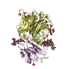
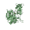
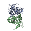
- Assembly
| Deposited unit | 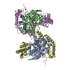
| ||||||||
|---|---|---|---|---|---|---|---|---|---|
| 1 | 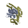
| ||||||||
| 2 | 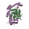
| ||||||||
| 3 | 
| ||||||||
| Unit cell |
| ||||||||
| Details | There are 2 biological units in the asymmetric unit (chains A & D and chains B & E) |
-Components
| #1: Protein | Mass: 33117.703 Da / Num. of mol.: 2 / Source method: isolated from a natural source / Source: (natural) Burkholderia glumae (bacteria) / Cellular location: extracellular / Strain: PG1References: UniProt: Q05489, UniProt: P0DUB8*PLUS, triacylglycerol lipase #2: Protein | Mass: 35236.238 Da / Num. of mol.: 2 / Fragment: periplasmic C-terminal domain Source method: isolated from a genetically manipulated source Source: (gene. exp.) Burkholderia glumae (bacteria) / Gene: lifO, lipB / Plasmid: pET16b / Production host: #3: Chemical |   Mass: 40.078 Da / Num. of mol.: 2 / Source method: obtained synthetically / Formula: Ca Mass: 40.078 Da / Num. of mol.: 2 / Source method: obtained synthetically / Formula: Ca#4: Chemical | ChemComp-IOD /   Mass: 126.904 Da / Num. of mol.: 8 / Source method: obtained synthetically / Formula: I Mass: 126.904 Da / Num. of mol.: 8 / Source method: obtained synthetically / Formula: I#5: Water | ChemComp-HOH / |  Mass: 18.015 Da / Num. of mol.: 843 / Source method: isolated from a natural source / Formula: H2O Mass: 18.015 Da / Num. of mol.: 843 / Source method: isolated from a natural source / Formula: H2OHas protein modification | Y | |
|---|
-Experimental details
-Experiment
| Experiment | Method: X-RAY DIFFRACTION / Number of used crystals: 1 |
|---|
- Sample preparation
| Crystal | Density Matthews: 2.6 Å3/Da / Density % sol: 52.9 % |
|---|---|
| Crystal grow | Temperature: 293 K / Method: vapor diffusion, hanging drop / pH: 8 Details: 20 % PEG3350, 0.2 M KI, pH 8.0, VAPOR DIFFUSION, HANGING DROP, temperature 293K |
-Data collection
| Diffraction | Mean temperature: 100 K |
|---|---|
| Diffraction source | Source: SYNCHROTRON / Site: ESRF  / Beamline: ID14-2 / Wavelength: 0.933 Å / Beamline: ID14-2 / Wavelength: 0.933 Å |
| Detector | Type: ADSC QUANTUM 4 / Detector: CCD |
| Radiation | Protocol: SINGLE WAVELENGTH / Monochromatic (M) / Laue (L): M / Scattering type: x-ray |
| Radiation wavelength | Wavelength: 0.933 Å / Relative weight: 1 |
| Reflection | Resolution: 1.85→40 Å / Num. all: 119154 / Num. obs: 120503 / % possible obs: 99 % / Redundancy: 7 % / Rmerge(I) obs: 0.072 / Χ2: 1.077 / Net I/σ(I): 15.4 |
| Reflection shell | Resolution: 1.85→1.92 Å / % possible obs: 91.3 % / Rmerge(I) obs: 0.521 / Mean I/σ(I) obs: 3.85 / Num. measured obs: 10920 / Χ2: 1.034 / % possible all: 91.3 |
- Processing
| Software |
| ||||||||||||||||||||||||
|---|---|---|---|---|---|---|---|---|---|---|---|---|---|---|---|---|---|---|---|---|---|---|---|---|---|
| Refinement | Method to determine structure: MOLECULAR REPLACEMENT Starting model: PDB ENTRY 1CVL Resolution: 1.85→40 Å
| ||||||||||||||||||||||||
| Displacement parameters | Biso mean: 34.93 Å2 | ||||||||||||||||||||||||
| Refinement step | Cycle: LAST / Resolution: 1.85→40 Å
|
