 Movie
Movie Controller

[English] 日本語
 Yorodumi
- PDB-1qge: NEW CRYSTAL FORM OF PSEUDOMONAS GLUMAE (FORMERLY CHROMOBACTERIUM ... -
+ Open data

- Basic information
| Entry | Database: PDB / ID: 1qge | ||||||
|---|---|---|---|---|---|---|---|
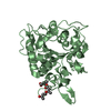
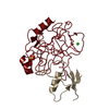
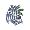
| Title | NEW CRYSTAL FORM OF PSEUDOMONAS GLUMAE (FORMERLY CHROMOBACTERIUM VISCOSUM ATCC 6918) LIPASE | ||||||
 Components Components | (PROTEIN (TRIACYLGLYCEROL HYDROLASE)) x 2 | ||||||
 Keywords Keywords | HYDROLASE / PSEUDOMONADACEAE / CIS-PEPTIDE / CLOSED CONFORMATION / LID | ||||||
| Function / homology |  Function and homology information Function and homology informationtriacylglycerol lipase / triacylglycerol lipase activity / lipid catabolic process / extracellular region / metal ion binding Similarity search - Function | ||||||
| Biological species |  Burkholderia glumae (bacteria) Burkholderia glumae (bacteria) | ||||||
| Method |  X-RAY DIFFRACTION / SYNCHROTRON / MOLECULAR REPLACEMENT / Resolution: 1.7 Å X-RAY DIFFRACTION / SYNCHROTRON / MOLECULAR REPLACEMENT / Resolution: 1.7 Å | ||||||
 Authors Authors | Lang, D.A. / Stadler, P. / Kovacs, A. / Paltauf, F. / Dijkstra, B.W. | ||||||
 Citation Citation | Journal: To be Published Title: Structural and Kinetic Investigations of Enantiomeric Binding Mode of Subclass I Lipases from the Family of Pseudomonadaceae Authors: Lang, D.A. / Stadler, P. / Kovacs, A. / Paltauf, F. / Dijkstra, B.W. #1: Journal: J.Mol.Biol. / Year: 1996Title: Crystal structure of a bacterial lipase from Chromobacterium viscosum ATCC 6918 refined at 1.6 angstroms resolution. Authors: Lang, D. / Hofmann, B. / Haalck, L. / Hecht, H.J. / Spener, F. / Schmid, R.D. / Schomburg, D. #2: Journal: Febs Lett. / Year: 1993Title: The crystal structure of triacylglycerol lipase from Pseudomonas glumae reveals a partially redundant catalytic aspartate. Authors: Noble, M.E. / Cleasby, A. / Johnson, L.N. / Egmond, M.R. / Frenken, L.G. | ||||||
| History |
|
- Structure visualization
| Structure viewer | Molecule: MolmilJmol/JSmol |
|---|
- Downloads & links
-Download
| PDBx/mmCIF format | 1qge.cif.gz | 77.9 KB | Display | PDBx/mmCIF format |
|---|---|---|---|---|
| PDB format | pdb1qge.ent.gz | 57.7 KB | Display | PDB format |
| PDBx/mmJSON format | 1qge.json.gz | Tree view | PDBx/mmJSON format | |
| Others |  Other downloads Other downloads |
-Validation report
| Arichive directory | https://data.pdbj.org/pub/pdb/validation_reports/qg/1qgeftp://data.pdbj.org/pub/pdb/validation_reports/qg/1qge | HTTPS FTP |
|---|
-Related structure data
| Related structure data |  1cvlS S: Starting model for refinement |
|---|---|
| Similar structure data |


-Links
 PDBj
PDBj


- Assembly
| Deposited unit | 
| ||||||||||
|---|---|---|---|---|---|---|---|---|---|---|---|
| 1 |
| ||||||||||
| Unit cell |
|
-Components
| #1: Protein | Mass: 22954.373 Da / Num. of mol.: 1 / Source method: isolated from a natural source / Details: ATCC / Source: (natural) Burkholderia glumae (bacteria) / Cellular location: EXTRACELLULAR / Strain: CHROMOBACTERIUM VISCOSUMReferences: UniProt: Q05489, UniProt: P0DUB9*PLUS, triacylglycerol lipase |
|---|---|
| #2: Protein | Mass: 10179.379 Da / Num. of mol.: 1 / Source method: isolated from a natural source / Details: ATCC / Source: (natural) Burkholderia glumae (bacteria) / Cellular location: EXTRACELLULAR / Strain: CHROMOBACTERIUM VISCOSUMReferences: UniProt: Q05489, UniProt: P0DUB9*PLUS, triacylglycerol lipase |
| #3: Chemical | ChemComp-CA /   Mass: 40.078 Da / Num. of mol.: 1 / Source method: obtained synthetically / Formula: Ca Mass: 40.078 Da / Num. of mol.: 1 / Source method: obtained synthetically / Formula: Ca |
| #4: Water | ChemComp-HOH /  Mass: 18.015 Da / Num. of mol.: 324 / Source method: isolated from a natural source / Formula: H2O Mass: 18.015 Da / Num. of mol.: 324 / Source method: isolated from a natural source / Formula: H2O |
| Compound details | PARTLY DEGRADED LIPASE AS A RESULT OF UNSPECIFIC PROTEOLYTIC DIGESTION DURING PURIFICATION AND/OR ...PARTLY DEGRADED LIPASE AS A RESULT OF UNSPECIFIC |
| Has protein modification | Y |
| Sequence details | 1QGE SWS Q05489 1 - 39 NOT IN ATOMS LIST REFERENCE: THE SEQUENCE FOR PSEUDOMONAS GLUMAE DESCRIBED ...1QGE SWS Q05489 1 - 39 NOT IN ATOMS LIST REFERENCE: THE SEQUENCE FOR PSEUDOMONA |
-Experimental details
-Experiment
| Experiment | Method: X-RAY DIFFRACTION / Number of used crystals: 1 |
|---|
- Sample preparation
| Crystal | Density Matthews: 1.9 Å3/Da / Density % sol: 35 % |
|---|---|
| Crystal grow | pH: 7.8 Details: PROTEIN WAS CRYSTALLIZED FROM 10 % PEG 6000, 5 % PEG 1000, 100 MM HEPES BUFFER, PH 7.8 |
-Data collection
| Diffraction | Mean temperature: 90 K |
|---|---|
| Diffraction source | Source: SYNCHROTRON / Site: MPG/DESY, HAMBURG  / Beamline: BW6 / Wavelength: 1 / Beamline: BW6 / Wavelength: 1 |
| Detector | Type: MARRESEARCH / Detector: IMAGE PLATE / Date: Dec 15, 1996 / Details: MIRRORS |
| Radiation | Monochromator: GRAPHITE / Protocol: SINGLE WAVELENGTH / Monochromatic (M) / Laue (L): M / Scattering type: x-ray |
| Radiation wavelength | Wavelength: 1 Å / Relative weight: 1 |
| Reflection | Resolution: 1.7→100 Å / Num. obs: 28124 / % possible obs: 98.6 % / Observed criterion σ(I): 2 / Redundancy: 3.4 % / Rsym value: 0.067 / Net I/σ(I): 10.5 |
| Reflection shell | Resolution: 1.7→1.73 Å / Redundancy: 2.5 % / Mean I/σ(I) obs: 4.1 / Rsym value: 0.27 / % possible all: 94.4 |
- Processing
| Software |
| ||||||||||||||||||||||||||||||||||||||||||||||||||||||||||||||||||||||||||||||||||||
|---|---|---|---|---|---|---|---|---|---|---|---|---|---|---|---|---|---|---|---|---|---|---|---|---|---|---|---|---|---|---|---|---|---|---|---|---|---|---|---|---|---|---|---|---|---|---|---|---|---|---|---|---|---|---|---|---|---|---|---|---|---|---|---|---|---|---|---|---|---|---|---|---|---|---|---|---|---|---|---|---|---|---|---|---|---|
| Refinement | Method to determine structure: MOLECULAR REPLACEMENT Starting model: PDB ENTRY 1CVL Resolution: 1.7→20 Å / Cross valid method: THROUGHOUT / σ(F): 0 Details: THE DISORDERED REGION (VAL 220, LEU 221 AND GLY 223) WAS NOT MODELED OR REFINED.
| ||||||||||||||||||||||||||||||||||||||||||||||||||||||||||||||||||||||||||||||||||||
| Displacement parameters | Biso mean: 14.9 Å2 | ||||||||||||||||||||||||||||||||||||||||||||||||||||||||||||||||||||||||||||||||||||
| Refinement step | Cycle: LAST / Resolution: 1.7→20 Å
| ||||||||||||||||||||||||||||||||||||||||||||||||||||||||||||||||||||||||||||||||||||
| Refine LS restraints |
|
