 Movie
Movie Controller

[English] 日本語
 Yorodumi
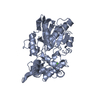
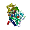
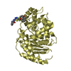
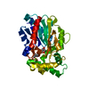
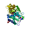
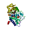
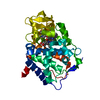
- PDB-1cvl: CRYSTAL STRUCTURE OF BACTERIAL LIPASE FROM CHROMOBACTERIUM VISCOS... -
+ Open data

- Basic information
| Entry | Database: PDB / ID: 1cvl | ||||||
|---|---|---|---|---|---|---|---|
| Title | CRYSTAL STRUCTURE OF BACTERIAL LIPASE FROM CHROMOBACTERIUM VISCOSUM ATCC 6918 | ||||||
 Components Components | TRIACYLGLYCEROL HYDROLASE | ||||||
 Keywords Keywords | HYDROLASE / TRIACYLGLYCEROL-HYDROLASE / PSEUDOMONADACEAE / OXYANION / CIS-PEPTIDE | ||||||
| Function / homology |  Function and homology information Function and homology informationtriacylglycerol lipase / triacylglycerol lipase activity / lipid catabolic process / extracellular region / metal ion binding Similarity search - Function | ||||||
| Biological species |  Chromobacterium viscosum (bacteria) Chromobacterium viscosum (bacteria) | ||||||
| Method |  X-RAY DIFFRACTION / SYNCHROTRON / MIR / Resolution: 1.6 Å X-RAY DIFFRACTION / SYNCHROTRON / MIR / Resolution: 1.6 Å | ||||||
 Authors Authors | Lang, D.A. / Hofmann, B. / Haalck, L. / Hecht, H.-J. / Spener, F. / Schmid, R.D. / Schomburg, D. | ||||||
 Citation Citation | Journal: J.Mol.Biol. / Year: 1996 Title: Crystal structure of a bacterial lipase from Chromobacterium viscosum ATCC 6918 refined at 1.6 angstroms resolution. Authors: Lang, D. / Hofmann, B. / Haalck, L. / Hecht, H.J. / Spener, F. / Schmid, R.D. / Schomburg, D. | ||||||
| History |
|
- Structure visualization
| Structure viewer | Molecule: MolmilJmol/JSmol |
|---|
- Downloads & links
-Download
| PDBx/mmCIF format | 1cvl.cif.gz | 72.2 KB | Display | PDBx/mmCIF format |
|---|---|---|---|---|
| PDB format | pdb1cvl.ent.gz | 53.7 KB | Display | PDB format |
| PDBx/mmJSON format | 1cvl.json.gz | Tree view | PDBx/mmJSON format | |
| Others |  Other downloads Other downloads |
-Validation report
| Arichive directory | https://data.pdbj.org/pub/pdb/validation_reports/cv/1cvlftp://data.pdbj.org/pub/pdb/validation_reports/cv/1cvl | HTTPS FTP |
|---|
-Related structure data
| Similar structure data |
|---|


-Links
 PDBj
PDBj


- Assembly
| Deposited unit | 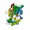
| ||||||||
|---|---|---|---|---|---|---|---|---|---|
| 1 |
| ||||||||
| Unit cell |
|
-Components
| #1: Protein | Mass: 33117.703 Da / Num. of mol.: 1 / Source method: isolated from a natural source / Details: CHAIN BREAK FROM V 220 - G 222 / Source: (natural) Chromobacterium viscosum (bacteria)References: UniProt: Q05489, UniProt: P0DUB9*PLUS, triacylglycerol lipase |
|---|---|
| #2: Chemical | ChemComp-CA /   Mass: 40.078 Da / Num. of mol.: 1 / Source method: obtained synthetically / Formula: Ca Mass: 40.078 Da / Num. of mol.: 1 / Source method: obtained synthetically / Formula: Ca |
| #3: Water | ChemComp-HOH /  Mass: 18.015 Da / Num. of mol.: 230 / Source method: isolated from a natural source / Formula: H2O Mass: 18.015 Da / Num. of mol.: 230 / Source method: isolated from a natural source / Formula: H2O |
| Compound details | PARTLY DEGRADED LIPASE AS A RESULT OF UNSPECIFIC PROTEOLYTIC DIGESTION DURING PURIFICATION AND/OR ...PARTLY DEGRADED LIPASE AS A RESULT OF UNSPECIFIC |
| Has protein modification | Y |
-Experimental details
-Experiment
| Experiment | Method: X-RAY DIFFRACTION / Number of used crystals: 1 |
|---|
- Sample preparation
| Crystal | Density Matthews: 2.15 Å3/Da / Density % sol: 42 % | |||||||||||||||||||||||||||||||||||
|---|---|---|---|---|---|---|---|---|---|---|---|---|---|---|---|---|---|---|---|---|---|---|---|---|---|---|---|---|---|---|---|---|---|---|---|---|
| Crystal grow | pH: 6.4 Details: PROTEIN WAS CRYSTALLIZED FROM 10-14 % PEG 4000, 10-14 % MPD, 100 MM CITRATE/PHOSPHATE BUFFER, PH 6.4 | |||||||||||||||||||||||||||||||||||
| Crystal grow | *PLUS Temperature: 292 K / Method: vapor diffusion, sitting drop | |||||||||||||||||||||||||||||||||||
| Components of the solutions | *PLUS
|
-Data collection
| Diffraction | Mean temperature: 293 K |
|---|---|
| Diffraction source | Source: SYNCHROTRON / Site: MPG/DESY, HAMBURG  / Beamline: BW6 / Wavelength: 1 / Beamline: BW6 / Wavelength: 1 |
| Detector | Type: MARRESEARCH / Detector: IMAGE PLATE / Date: Nov 1, 1993 / Details: MIRRORS |
| Radiation | Monochromator: GRAPHITE(002) / Monochromatic (M) / Laue (L): M / Scattering type: x-ray |
| Radiation wavelength | Wavelength: 1 Å / Relative weight: 1 |
| Reflection | Resolution: 1.6→20 Å / Num. obs: 33644 / % possible obs: 88 % / Observed criterion σ(I): 2 / Redundancy: 2.6 % / Rmerge(I) obs: 0.053 / Rsym value: 0.053 / Net I/σ(I): 8.5 |
| Reflection shell | Resolution: 1.6→1.64 Å / Redundancy: 1.2 % / Rmerge(I) obs: 0.053 / Mean I/σ(I) obs: 3.1 / Rsym value: 0.22 / % possible all: 73.7 |
| Reflection | *PLUS Num. measured all: 87722 |
| Reflection shell | *PLUS % possible obs: 73.7 % / Rmerge(I) obs: 0.22 |
- Processing
| Software |
| ||||||||||||||||||||||||||||||||||||||||||||||||||||||||||||||||||||||||||||||||||||
|---|---|---|---|---|---|---|---|---|---|---|---|---|---|---|---|---|---|---|---|---|---|---|---|---|---|---|---|---|---|---|---|---|---|---|---|---|---|---|---|---|---|---|---|---|---|---|---|---|---|---|---|---|---|---|---|---|---|---|---|---|---|---|---|---|---|---|---|---|---|---|---|---|---|---|---|---|---|---|---|---|---|---|---|---|---|
| Refinement | Method to determine structure: MIR / Resolution: 1.6→8 Å / Cross valid method: RANDOM / σ(F): 0
| ||||||||||||||||||||||||||||||||||||||||||||||||||||||||||||||||||||||||||||||||||||
| Displacement parameters | Biso mean: 14.9 Å2 | ||||||||||||||||||||||||||||||||||||||||||||||||||||||||||||||||||||||||||||||||||||
| Refine analyze | Luzzati coordinate error obs: 0.18 Å / Luzzati d res low obs: 8 Å / Luzzati sigma a obs: 0.18 Å | ||||||||||||||||||||||||||||||||||||||||||||||||||||||||||||||||||||||||||||||||||||
| Refinement step | Cycle: LAST / Resolution: 1.6→8 Å
| ||||||||||||||||||||||||||||||||||||||||||||||||||||||||||||||||||||||||||||||||||||
| Refine LS restraints |
| ||||||||||||||||||||||||||||||||||||||||||||||||||||||||||||||||||||||||||||||||||||
| Software | *PLUS Name: PROLSQ / Classification: refinement | ||||||||||||||||||||||||||||||||||||||||||||||||||||||||||||||||||||||||||||||||||||
| Refinement | *PLUS Rfactor obs: 0.178 | ||||||||||||||||||||||||||||||||||||||||||||||||||||||||||||||||||||||||||||||||||||
| Solvent computation | *PLUS | ||||||||||||||||||||||||||||||||||||||||||||||||||||||||||||||||||||||||||||||||||||
| Displacement parameters | *PLUS |
