 Movie
Movie Controller

[English] 日本語
 Yorodumi
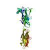
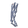
- PDB-5u5o: Bacterial adhesin from Mobiluncus mulieris containing intramolecu... -
+ Open data

- Basic information
| Entry | Database: PDB / ID: 5u5o | ||||||
|---|---|---|---|---|---|---|---|
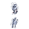
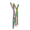
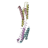
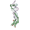
| Title | Bacterial adhesin from Mobiluncus mulieris containing intramolecular disulfide, isopeptide, and ester bond cross-links (space group P1) | ||||||
 Components Components | LPXTG-motif cell wall anchor domain protein | ||||||
 Keywords Keywords | CELL ADHESION / bacterial adhesin / Ig-like domain / intramolecular cross-link | ||||||
| Function / homology | Domain of unknown function DUF5979 / T-Q ester bond containing domain / T-Q ester bond containing domain / Domain of unknown function (DUF5979) / membrane / LPXTG-motif cell wall anchor domain protein Function and homology information Function and homology information | ||||||
| Biological species |  Mobiluncus mulieris (bacteria) Mobiluncus mulieris (bacteria) | ||||||
| Method |  X-RAY DIFFRACTION / SYNCHROTRON / MOLECULAR REPLACEMENT / Resolution: 1.15 Å X-RAY DIFFRACTION / SYNCHROTRON / MOLECULAR REPLACEMENT / Resolution: 1.15 Å | ||||||
 Authors Authors | Paynter, J. / Young, P.G. / Squire, C.J. | ||||||
| Funding support |  New Zealand, 1items New Zealand, 1items
| ||||||
 Citation Citation | Journal: Acta Crystallogr D Struct Biol / Year: 2023 Title: Domain structure and cross-linking in a giant adhesin from the Mobiluncus mulieris bacterium. Authors: Young, P.G. / Paynter, J.M. / Wardega, J.K. / Middleditch, M.J. / Payne, L.S. / Baker, E.N. / Squire, C.J. | ||||||
| History |
|
- Structure visualization
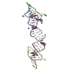
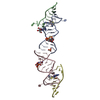
| Structure viewer | Molecule: MolmilJmol/JSmol |
|---|
- Downloads & links
-Download
| PDBx/mmCIF format | 5u5o.cif.gz | 138.6 KB | Display | PDBx/mmCIF format |
|---|---|---|---|---|
| PDB format | pdb5u5o.ent.gz | 108.3 KB | Display | PDB format |
| PDBx/mmJSON format | 5u5o.json.gz | Tree view | PDBx/mmJSON format | |
| Others |  Other downloads Other downloads |
-Validation report
| Summary document | 5u5o_validation.pdf.gz | 413.8 KB | Display | wwPDB validaton report |
|---|---|---|---|---|
| Full document | 5u5o_full_validation.pdf.gz | 415.2 KB | Display | |
| Data in XML | 5u5o_validation.xml.gz | 15 KB | Display | |
| Data in CIF | 5u5o_validation.cif.gz | 23 KB | Display | |
| Arichive directory | https://data.pdbj.org/pub/pdb/validation_reports/u5/5u5oftp://data.pdbj.org/pub/pdb/validation_reports/u5/5u5o | HTTPS FTP |
-Related structure data


-Links
 PDBj
PDBj- Assembly
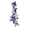
| Deposited unit | 
| ||||||||
|---|---|---|---|---|---|---|---|---|---|
| 1 |
| ||||||||
| Unit cell |
|
-Components
| #1: Protein | Mass: 31388.865 Da / Num. of mol.: 1 / Fragment: UNP residues 6668-6959 Source method: isolated from a genetically manipulated source Source: (gene. exp.) Mobiluncus mulieris (bacteria) / Plasmid: pPROEX-Hta / Production host: |
|---|---|
| #2: Water | ChemComp-HOH /  Mass: 18.015 Da / Num. of mol.: 326 / Source method: isolated from a natural source / Formula: H2O Mass: 18.015 Da / Num. of mol.: 326 / Source method: isolated from a natural source / Formula: H2O |
| Has protein modification | Y |
-Experimental details
-Experiment
| Experiment | Method: X-RAY DIFFRACTION / Number of used crystals: 1 |
|---|
- Sample preparation
| Crystal | Density Matthews: 2.52 Å3/Da / Density % sol: 51.19 % |
|---|---|
| Crystal grow | Temperature: 291 K / Method: vapor diffusion, hanging drop / pH: 6.5 Details: 10% (v/v) PEG 8000, 20% (v/v) ethylene glycol, 0.02 M carboxylic acids (sodium formate, ammonium acetate, trisodium citrate, sodium potassium tartrate, sodium oxamate), and 0.1 M MES/Imidazole (pH 6.5). |
-Data collection
| Diffraction | Mean temperature: 100 K |
|---|---|
| Diffraction source | Source: SYNCHROTRON / Site: Australian Synchrotron  / Beamline: MX1 / Wavelength: 0.9537 Å / Beamline: MX1 / Wavelength: 0.9537 Å |
| Detector | Type: ADSC QUANTUM 210r / Detector: CCD / Date: Oct 25, 2015 |
| Radiation | Protocol: SINGLE WAVELENGTH / Monochromatic (M) / Laue (L): M / Scattering type: x-ray |
| Radiation wavelength | Wavelength: 0.9537 Å / Relative weight: 1 |
| Reflection | Resolution: 1.15→19.15 Å / Num. obs: 102468 / % possible obs: 94.1 % / Redundancy: 7.9 % / CC1/2: 1 / Net I/σ(I): 20.2 |
| Reflection shell | Resolution: 1.15→1.17 Å / Redundancy: 7.9 % / Mean I/σ(I) obs: 1 / Num. measured obs: 4921 / CC1/2: 0.549 / % possible all: 90.6 |
- Processing
| Software |
| ||||||||||||||||||||||||||||||||||||||||||||||||||||||||||||||||||||||||||||||||||||||||||||||||||||||||||||||||||||||||||||||||||||||||||||||||||||||||||||||||||||||||||||||||||||||
|---|---|---|---|---|---|---|---|---|---|---|---|---|---|---|---|---|---|---|---|---|---|---|---|---|---|---|---|---|---|---|---|---|---|---|---|---|---|---|---|---|---|---|---|---|---|---|---|---|---|---|---|---|---|---|---|---|---|---|---|---|---|---|---|---|---|---|---|---|---|---|---|---|---|---|---|---|---|---|---|---|---|---|---|---|---|---|---|---|---|---|---|---|---|---|---|---|---|---|---|---|---|---|---|---|---|---|---|---|---|---|---|---|---|---|---|---|---|---|---|---|---|---|---|---|---|---|---|---|---|---|---|---|---|---|---|---|---|---|---|---|---|---|---|---|---|---|---|---|---|---|---|---|---|---|---|---|---|---|---|---|---|---|---|---|---|---|---|---|---|---|---|---|---|---|---|---|---|---|---|---|---|---|---|
| Refinement | Method to determine structure: MOLECULAR REPLACEMENT Starting model: in house model Resolution: 1.15→19.15 Å / Cor.coef. Fo:Fc: 0.972 / Cor.coef. Fo:Fc free: 0.963 / SU B: 2.376 / SU ML: 0.046 / Cross valid method: THROUGHOUT / ESU R: 0.04 / ESU R Free: 0.041 / Details: HYDROGENS HAVE BEEN ADDED IN THE RIDING POSITIONS
| ||||||||||||||||||||||||||||||||||||||||||||||||||||||||||||||||||||||||||||||||||||||||||||||||||||||||||||||||||||||||||||||||||||||||||||||||||||||||||||||||||||||||||||||||||||||
| Solvent computation | Ion probe radii: 0.8 Å / Shrinkage radii: 0.8 Å / VDW probe radii: 1.2 Å | ||||||||||||||||||||||||||||||||||||||||||||||||||||||||||||||||||||||||||||||||||||||||||||||||||||||||||||||||||||||||||||||||||||||||||||||||||||||||||||||||||||||||||||||||||||||
| Displacement parameters | Biso mean: 18.59 Å2
| ||||||||||||||||||||||||||||||||||||||||||||||||||||||||||||||||||||||||||||||||||||||||||||||||||||||||||||||||||||||||||||||||||||||||||||||||||||||||||||||||||||||||||||||||||||||
| Refinement step | Cycle: 1 / Resolution: 1.15→19.15 Å
| ||||||||||||||||||||||||||||||||||||||||||||||||||||||||||||||||||||||||||||||||||||||||||||||||||||||||||||||||||||||||||||||||||||||||||||||||||||||||||||||||||||||||||||||||||||||
| Refine LS restraints |
|