 Movie
Movie Controller

[English] 日本語
 Yorodumi
- PDB-1ovo: CRYSTALLOGRAPHIC REFINEMENT OF JAPANESE QUAIL OVOMUCOID, A KAZAL-... -
+ Open data

- Basic information
| Entry | Database: PDB / ID: 1ovo | ||||||
|---|---|---|---|---|---|---|---|
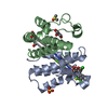
| Title | CRYSTALLOGRAPHIC REFINEMENT OF JAPANESE QUAIL OVOMUCOID, A KAZAL-TYPE INHIBITOR, AND MODEL BUILDING STUDIES OF COMPLEXES WITH SERINE PROTEASES | ||||||
 Components Components | OVOMUCOID THIRD DOMAIN | ||||||
 Keywords Keywords | PROTEINASE INHIBITOR (KAZAL) | ||||||
| Function / homology |  Function and homology information Function and homology information | ||||||
| Biological species |  Coturnix japonica (Japanese quail) Coturnix japonica (Japanese quail) | ||||||
| Method |  X-RAY DIFFRACTION / Resolution: 1.9 Å X-RAY DIFFRACTION / Resolution: 1.9 Å | ||||||
 Authors Authors | Weber, E. / Papamokos, E. / Bode, W. / Huber, R. / Kato, I. / Laskowskijunior, M. | ||||||
 Citation Citation | Journal: J.Mol.Biol. / Year: 1982 Title: Crystallographic refinement of Japanese quail ovomucoid, a Kazal-type inhibitor, and model building studies of complexes with serine proteases. Authors: Papamokos, E. / Weber, E. / Bode, W. / Huber, R. / Empie, M.W. / Kato, I. / Laskowski Jr., M. #1: Journal: Acta Crystallogr.,Sect.B / Year: 1983Title: The Geometry of the Reactive Site and of the Peptide Groups in Trypsin, Trypsinogen and its Complexes with Inhibitors Authors: Marquart, M. / Walter, J. / Deisenhofer, J. / Bode, W. / Huber, R. #2: Journal: J.Mol.Biol. / Year: 1981Title: Crystallization,Crystal Structure Analysis and Molecular Model of the Third Domain of Japanese Quail Ovomucoid,A Kazal Type Inhibitor Authors: Weber, E. / Papamokos, E. / Bode, W. / Huber, R. / Kato, I. / Laskowskijunior, M. | ||||||
| History |
|
- Structure visualization
| Structure viewer | Molecule: MolmilJmol/JSmol |
|---|
- Downloads & links
-Download
| PDBx/mmCIF format | 1ovo.cif.gz | 49.5 KB | Display | PDBx/mmCIF format |
|---|---|---|---|---|
| PDB format | pdb1ovo.ent.gz | 38.2 KB | Display | PDB format |
| PDBx/mmJSON format | 1ovo.json.gz | Tree view | PDBx/mmJSON format | |
| Others |  Other downloads Other downloads |
-Validation report
| Arichive directory | https://data.pdbj.org/pub/pdb/validation_reports/ov/1ovoftp://data.pdbj.org/pub/pdb/validation_reports/ov/1ovo | HTTPS FTP |
|---|
-Related structure data
| Similar structure data |
|---|


-Links
 PDBj
PDBj
- Assembly
| Deposited unit | 
| ||||||||||||
|---|---|---|---|---|---|---|---|---|---|---|---|---|---|
| 1 | 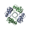
| ||||||||||||
| 2 | 
| ||||||||||||
| 3 | 
| ||||||||||||
| Unit cell |
| ||||||||||||
| Atom site foot note | 1: SEE REMARK 5. / 2: RESIDUE 12 OF EACH CHAIN IS A CIS-PROLINE. | ||||||||||||
| Noncrystallographic symmetry (NCS) | NCS oper:
| ||||||||||||
| Details | THE MTRIX RECORDS BELOW DESCRIBE THE NON-CRYSTALLOGRAPHIC RELATIONSHIPS AMONG THE INDIVIDUAL DOMAINS IN THIS ENTRY. MTRIX 1 RELATES CHAIN A TO CHAIN B BY A PSEUDO-FOUR-FOLD ROTATION. MTRIX 2 RELATES CHAIN C TO CHAIN D BY A PSEUDO-TWO-FOLD ROTATION. SEE REFERENCE 2 ABOVE FOR A COMPLETE DESCRIPTION OF THESE SYMMETRY AXES. THESE MATRICES WERE OBTAINED BY COMPARING THE BACKBONE AND CB ATOMS OF THE MOLECULES. (THE FIRST SIX RESIDUES WERE EXCLUDED FROM THIS CALCULATION.) |
-Components
| #1: Protein | Mass: 6067.842 Da / Num. of mol.: 4 Source method: isolated from a genetically manipulated source Source: (gene. exp.) Coturnix japonica (Japanese quail) / References: UniProt: P01003#2: Water | ChemComp-HOH / |  Mass: 18.015 Da / Num. of mol.: 46 / Source method: isolated from a natural source / Formula: H2O Mass: 18.015 Da / Num. of mol.: 46 / Source method: isolated from a natural source / Formula: H2OHas protein modification | Y | Sequence details | THE MATERIAL USED IS POLYMORPHIC WITH 20 PER CENT GLY 32 AND 80 PER CENT SER 32. IT IS PREMATURE AT ...THE MATERIAL USED IS POLYMORPHI | |
|---|
-Experimental details
-Experiment
| Experiment | Method: X-RAY DIFFRACTION |
|---|
- Sample preparation
| Crystal | Density Matthews: 2.79 Å3/Da / Density % sol: 55.88 % |
|---|---|
| Crystal grow | *PLUS Temperature: 20 ℃ / pH: 9.5 / Method: unknown / Details: Weber, E., (1981) J.Mol.Biol., 149, 109. |
| Components of the solutions | *PLUS Conc.: 0.96-1.0 M / Common name: citrate |
-Data collection
| Radiation | Scattering type: x-ray |
|---|---|
| Radiation wavelength | Relative weight: 1 |
| Reflection | *PLUS Highest resolution: 1.9 Å / Num. obs: 19537 / % possible obs: 75 % / Num. measured all: 63795 / Rmerge(I) obs: 0.084 |
- Processing
| Software | Name: REAL-SPACE / Version: REFINEMENT / Classification: refinement | ||||||||||||
|---|---|---|---|---|---|---|---|---|---|---|---|---|---|
| Refinement | Highest resolution: 1.9 Å Details: AN OCCUPANCY OF 0.0 INDICATES THAT NO SIGNIFICANT ELECTRON DENSITY WAS FOUND IN THE FINAL FOURIER MAP. | ||||||||||||
| Refinement step | Cycle: LAST / Highest resolution: 1.9 Å
| ||||||||||||
| Refinement | *PLUS Lowest resolution: 6 Å / Num. reflection obs: 15980 / Rfactor obs: 0.207 | ||||||||||||
| Solvent computation | *PLUS | ||||||||||||
| Displacement parameters | *PLUS |
