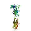
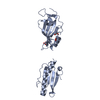
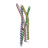
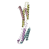
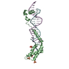
[English] 日本語
Yorodumi
- PDB-5u5o: Bacterial adhesin from Mobiluncus mulieris containing intramolecu... -

+
Open data


ID or keywords:

Loading...

-
Basic information

Entry
Database: PDB / ID: 5u5o
TitleBacterial adhesin from Mobiluncus mulieris containing intramolecular disulfide, isopeptide, and ester bond cross-links (space group P1)
ComponentsLPXTG-motif cell wall anchor domain protein
KeywordsCELL ADHESION / bacterial adhesin / Ig-like domain / intramolecular cross-link
Function / homologyT-Q ester bond containing domain / T-Q ester bond containing domain / Domain of unknown function DUF5979 / Domain of unknown function (DUF5979) / membrane / LPXTG-motif cell wall anchor domain protein
Function and homology information
Biological speciesMobiluncus mulieris (bacteria)
MethodX-RAY DIFFRACTION / SYNCHROTRON / MOLECULAR REPLACEMENT / Resolution: 1.15 Å
AuthorsPaynter, J. / Young, P.G. / Squire, C.J.
Funding support New Zealand, 1items
OrganizationGrant numberCountry
Royal Society of NZ Marsden Fund New Zealand
CitationJournal: Acta Crystallogr D Struct Biol / Year: 2023
Title: Domain structure and cross-linking in a giant adhesin from the Mobiluncus mulieris bacterium.
Authors: Young, P.G. / Paynter, J.M. / Wardega, J.K. / Middleditch, M.J. / Payne, L.S. / Baker, E.N. / Squire, C.J.
History
DepositionDec 7, 2016Deposition site: RCSB / Processing site: RCSB
Revision 1.0Jun 13, 2018Provider: repository / Type: Initial release
Revision 1.1Apr 3, 2024Group: Data collection / Database references / Refinement description
Category: chem_comp_atom / chem_comp_bond ...chem_comp_atom / chem_comp_bond / citation / citation_author / database_2 / pdbx_initial_refinement_model
Item: _citation.journal_abbrev / _citation.journal_id_CSD ..._citation.journal_abbrev / _citation.journal_id_CSD / _citation.journal_id_ISSN / _citation.journal_volume / _citation.page_first / _citation.page_last / _citation.pdbx_database_id_DOI / _citation.pdbx_database_id_PubMed / _citation.title / _citation.year / _database_2.pdbx_DOI / _database_2.pdbx_database_accession

-
Structure visualization

Structure viewerMolecule:
MolmilJmol/JSmol

Downloads & links

-
Assembly

Deposited unit
A: LPXTG-motif cell wall anchor domain protein


Theoretical massNumber of molelcules
Total (without water)31,3891
Polymers31,3891
Non-polymers00
Water5,873326
1


  • Idetical with deposited unit
  • defined by author&software
TypeNameSymmetry operationNumber
identity operation1_555x,y,z1
MethodPISA
Unit cell
Length a, b, c (Å)27.900, 54.983, 57.317
Angle α, β, γ (deg.)67.72, 76.47, 85.35
Int Tables number1
Space group name H-MP1

-
Components

#1: Protein LPXTG-motif cell wall anchor domain protein


Mass: 31388.865 Da / Num. of mol.: 1 / Fragment: UNP residues 6668-6959
Source method: isolated from a genetically manipulated source
Source: (gene. exp.) Mobiluncus mulieris (bacteria) / Plasmid: pPROEX-Hta / Production host: Escherichia coli (E. coli) / References: UniProt: E0QN07
#2: Water ChemComp-HOH / water


Mass: 18.015 Da / Num. of mol.: 326 / Source method: isolated from a natural source / Formula: H2O

-
Experimental details

-
Experiment

ExperimentMethod: X-RAY DIFFRACTION / Number of used crystals: 1

-
Sample preparation

CrystalDensity Matthews: 2.52 Å3/Da / Density % sol: 51.19 %
Crystal growTemperature: 291 K / Method: vapor diffusion, hanging drop / pH: 6.5
Details: 10% (v/v) PEG 8000, 20% (v/v) ethylene glycol, 0.02 M carboxylic acids (sodium formate, ammonium acetate, trisodium citrate, sodium potassium tartrate, sodium oxamate), and 0.1 M MES/Imidazole (pH 6.5).

-
Data collection

DiffractionMean temperature: 100 K
Diffraction sourceSource: SYNCHROTRON / Site: Australian Synchrotron / Beamline: MX1 / Wavelength: 0.9537 Å
DetectorType: ADSC QUANTUM 210r / Detector: CCD / Date: Oct 25, 2015
RadiationProtocol: SINGLE WAVELENGTH / Monochromatic (M) / Laue (L): M / Scattering type: x-ray
Radiation wavelengthWavelength: 0.9537 Å / Relative weight: 1
ReflectionResolution: 1.15→19.15 Å / Num. obs: 102468 / % possible obs: 94.1 % / Redundancy: 7.9 % / CC1/2: 1 / Net I/σ(I): 20.2
Reflection shellResolution: 1.15→1.17 Å / Redundancy: 7.9 % / Mean I/σ(I) obs: 1 / Num. measured obs: 4921 / CC1/2: 0.549 / % possible all: 90.6

-
Processing

Software
NameVersionClassification
REFMAC5.8.0103refinement
XDSdata reduction
Aimlessdata scaling
PHASERphasing
RefinementMethod to determine structure: MOLECULAR REPLACEMENT
Starting model: in house model

Resolution: 1.15→19.15 Å / Cor.coef. Fo:Fc: 0.972 / Cor.coef. Fo:Fc free: 0.963 / SU B: 2.376 / SU ML: 0.046 / Cross valid method: THROUGHOUT / ESU R: 0.04 / ESU R Free: 0.041 / Details: HYDROGENS HAVE BEEN ADDED IN THE RIDING POSITIONS
RfactorNum. reflection% reflectionSelection details
Rfree0.21054 5177 5.1 %RANDOM
Rwork0.18141 ---
obs0.18289 97278 94.02 %-
Solvent computationIon probe radii: 0.8 Å / Shrinkage radii: 0.8 Å / VDW probe radii: 1.2 Å
Displacement parametersBiso mean: 18.59 Å2
Baniso -1Baniso -2Baniso -3
1--0.04 Å2-0.02 Å2-0.03 Å2
2---0 Å2-0 Å2
3---0.01 Å2
Refinement stepCycle: 1 / Resolution: 1.15→19.15 Å
ProteinNucleic acidLigandSolventTotal
Num. atoms2160 0 0 326 2486
Refine LS restraints
Refine-IDTypeDev idealDev ideal targetNumber
X-RAY DIFFRACTIONr_bond_refined_d0.0110.022276
X-RAY DIFFRACTIONr_bond_other_d0.0020.022150
X-RAY DIFFRACTIONr_angle_refined_deg1.4821.9613101
X-RAY DIFFRACTIONr_angle_other_deg0.84234999
X-RAY DIFFRACTIONr_dihedral_angle_1_deg5.9485.033307
X-RAY DIFFRACTIONr_dihedral_angle_2_deg34.62526.96689
X-RAY DIFFRACTIONr_dihedral_angle_3_deg10.5115398
X-RAY DIFFRACTIONr_dihedral_angle_4_deg12.08153
X-RAY DIFFRACTIONr_chiral_restr0.0850.2370
X-RAY DIFFRACTIONr_gen_planes_refined0.0060.022615
X-RAY DIFFRACTIONr_gen_planes_other0.0010.02442
X-RAY DIFFRACTIONr_nbd_refined
X-RAY DIFFRACTIONr_nbd_other
X-RAY DIFFRACTIONr_nbtor_refined
X-RAY DIFFRACTIONr_nbtor_other
X-RAY DIFFRACTIONr_xyhbond_nbd_refined
X-RAY DIFFRACTIONr_xyhbond_nbd_other
X-RAY DIFFRACTIONr_metal_ion_refined
X-RAY DIFFRACTIONr_metal_ion_other
X-RAY DIFFRACTIONr_symmetry_vdw_refined
X-RAY DIFFRACTIONr_symmetry_vdw_other
X-RAY DIFFRACTIONr_symmetry_hbond_refined
X-RAY DIFFRACTIONr_symmetry_hbond_other
X-RAY DIFFRACTIONr_symmetry_metal_ion_refined
X-RAY DIFFRACTIONr_symmetry_metal_ion_other
X-RAY DIFFRACTIONr_mcbond_it1.8581.6621194
X-RAY DIFFRACTIONr_mcbond_other1.8581.6611193
X-RAY DIFFRACTIONr_mcangle_it2.472.5061504
X-RAY DIFFRACTIONr_mcangle_other2.4692.5071505
X-RAY DIFFRACTIONr_scbond_it2.0691.8861082
X-RAY DIFFRACTIONr_scbond_other2.0681.8861082
X-RAY DIFFRACTIONr_scangle_it
X-RAY DIFFRACTIONr_scangle_other2.5752.7241595
X-RAY DIFFRACTIONr_long_range_B_refined3.63115.3322658
X-RAY DIFFRACTIONr_long_range_B_other3.39614.3042487
X-RAY DIFFRACTIONr_rigid_bond_restr1.92834426
X-RAY DIFFRACTIONr_sphericity_free28.253562
X-RAY DIFFRACTIONr_sphericity_bonded15.49154647
LS refinement shellResolution: 1.15→1.18 Å / Total num. of bins used: 20
RfactorNum. reflection% reflection
Rfree0.286 375 -
Rwork0.281 6942 -
obs--90.57 %

+
About Yorodumi

-
News

-
Feb 9, 2022. New format data for meta-information of EMDB entries

New format data for meta-information of EMDB entries

  • Version 3 of the EMDB header file is now the official format.
  • The previous official version 1.9 will be removed from the archive.

Related info.:EMDB header

External links:wwPDB to switch to version 3 of the EMDB data model

-
Aug 12, 2020. Covid-19 info

Covid-19 info

URL: https://pdbjlvh1.pdbj.org/emnavi/covid19.php

New page: Covid-19 featured information page in EM Navigator.

Related info.:Covid-19 info / Mar 5, 2020. Novel coronavirus structure data

+
Mar 5, 2020. Novel coronavirus structure data

Novel coronavirus structure data

Related info.:Yorodumi Speices / Aug 12, 2020. Covid-19 info

External links:COVID-19 featured content - PDBj / Molecule of the Month (242):Coronavirus Proteases

+
Jan 31, 2019. EMDB accession codes are about to change! (news from PDBe EMDB page)

EMDB accession codes are about to change! (news from PDBe EMDB page)

  • The allocation of 4 digits for EMDB accession codes will soon come to an end. Whilst these codes will remain in use, new EMDB accession codes will include an additional digit and will expand incrementally as the available range of codes is exhausted. The current 4-digit format prefixed with “EMD-” (i.e. EMD-XXXX) will advance to a 5-digit format (i.e. EMD-XXXXX), and so on. It is currently estimated that the 4-digit codes will be depleted around Spring 2019, at which point the 5-digit format will come into force.
  • The EM Navigator/Yorodumi systems omit the EMD- prefix.

Related info.:Q: What is EMD? / ID/Accession-code notation in Yorodumi/EM Navigator

External links:EMDB Accession Codes are Changing Soon! / Contact to PDBj

+
Jul 12, 2017. Major update of PDB

Major update of PDB

  • wwPDB released updated PDB data conforming to the new PDBx/mmCIF dictionary.
  • This is a major update changing the version number from 4 to 5, and with Remediation, in which all the entries are updated.
  • In this update, many items about electron microscopy experimental information are reorganized (e.g. em_software).
  • Now, EM Navigator and Yorodumi are based on the updated data.

External links:wwPDB Remediation / Enriched Model Files Conforming to OneDep Data Standards Now Available in the PDB FTP Archive

-
Yorodumi

Thousand views of thousand structures

  • Yorodumi is a browser for structure data from EMDB, PDB, SASBDB, etc.
  • This page is also the successor to EM Navigator detail page, and also detail information page/front-end page for Omokage search.
  • The word "yorodu" (or yorozu) is an old Japanese word meaning "ten thousand". "mi" (miru) is to see.

Related info.:EMDB / PDB / SASBDB / Comparison of 3 databanks / Yorodumi Search / Aug 31, 2016. New EM Navigator & Yorodumi / Yorodumi Papers / Jmol/JSmol / Function and homology information / Changes in new EM Navigator and Yorodumi

Read more