 Movie
Movie Controller

[English] 日本語
 Yorodumi
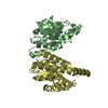
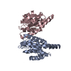
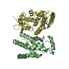
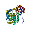
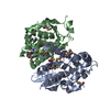
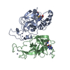
- PDB-5u51: Structure of Francisella tularensis heterodimeric SspA (MglA-SspA... -
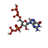
+ Open data

- Basic information
| Entry | Database: PDB / ID: 5u51 | ||||||
|---|---|---|---|---|---|---|---|
| Title | Structure of Francisella tularensis heterodimeric SspA (MglA-SspA) in complex with ppGpp | ||||||
 Components Components |
| ||||||
 Keywords Keywords | TRANSCRIPTION / Stringent starvation protein A / Macrophage growth locus A / ppGpp / GST-fold | ||||||
| Function / homology |  Function and homology information Function and homology information | ||||||
| Biological species |  Francisella tularensis (bacteria) Francisella tularensis (bacteria) | ||||||
| Method |  X-RAY DIFFRACTION / SYNCHROTRON / MOLECULAR REPLACEMENT / Resolution: 2.8 Å X-RAY DIFFRACTION / SYNCHROTRON / MOLECULAR REPLACEMENT / Resolution: 2.8 Å | ||||||
 Authors Authors | Cuthbert, B.J. / Schumacher, M.A. / Brennan, R.G. | ||||||
 Citation Citation | Journal: Genes Dev. / Year: 2017 Title: Dissection of the molecular circuitry controlling virulence in Francisella tularensis. Authors: Cuthbert, B.J. / Ross, W. / Rohlfing, A.E. / Dove, S.L. / Gourse, R.L. / Brennan, R.G. / Schumacher, M.A. | ||||||
| History |
|
- Structure visualization
| Structure viewer | Molecule: MolmilJmol/JSmol |
|---|
- Downloads & links
-Download
| PDBx/mmCIF format | 5u51.cif.gz | 176.5 KB | Display | PDBx/mmCIF format |
|---|---|---|---|---|
| PDB format | pdb5u51.ent.gz | 136.4 KB | Display | PDB format |
| PDBx/mmJSON format | 5u51.json.gz | Tree view | PDBx/mmJSON format | |
| Others |  Other downloads Other downloads |
-Validation report
| Arichive directory | https://data.pdbj.org/pub/pdb/validation_reports/u5/5u51ftp://data.pdbj.org/pub/pdb/validation_reports/u5/5u51 | HTTPS FTP |
|---|
-Related structure data
| Related structure data | 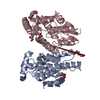 5u56C  6alxC  1yy7S  4purS C: citing same article ( S: Starting model for refinement |
|---|---|
| Similar structure data |


-Links
 PDBj
PDBj

- Assembly
| Deposited unit | 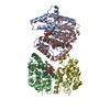
| ||||||||
|---|---|---|---|---|---|---|---|---|---|
| 1 | 
| ||||||||
| 2 | 
| ||||||||
| Unit cell |
|
-Components
-Protein , 2 types, 4 molecules CDAB
| #1: Protein | Mass: 24110.094 Da / Num. of mol.: 2 Source method: isolated from a genetically manipulated source Source: (gene. exp.) Francisella tularensis (bacteria) / Gene: DR86_1150 / Plasmid: pMCSG21 / Details (production host): For coexpression / Cell line (production host): C41 (DE3) / Production host: #2: Protein | Mass: 23594.529 Da / Num. of mol.: 2 Source method: isolated from a genetically manipulated source Source: (gene. exp.) Francisella tularensis (bacteria) / Gene: DR86_1530 / Plasmid: pET28ADetails (production host): Expressed as an MBP fusion from pMCSG9 Cell line (production host): C41 (DE3) / Production host: |
|---|
-Non-polymers , 5 types, 54 molecules 


| #3: Chemical | ChemComp-GOL /  Mass: 92.094 Da / Num. of mol.: 5 / Source method: obtained synthetically / Formula: C3H8O3 Mass: 92.094 Da / Num. of mol.: 5 / Source method: obtained synthetically / Formula: C3H8O3#4: Chemical |  Mass: 238.278 Da / Num. of mol.: 2 / Source method: obtained synthetically / Formula: C10H22O6 / Comment: precipitant*YM Mass: 238.278 Da / Num. of mol.: 2 / Source method: obtained synthetically / Formula: C10H22O6 / Comment: precipitant*YM#5: Chemical |  Mass: 24.305 Da / Num. of mol.: 2 / Source method: obtained synthetically / Formula: Mg Mass: 24.305 Da / Num. of mol.: 2 / Source method: obtained synthetically / Formula: Mg#6: Chemical |  Type: RNA linking / Mass: 603.160 Da / Num. of mol.: 2 / Source method: obtained synthetically / Formula: C10H17N5O17P4 Type: RNA linking / Mass: 603.160 Da / Num. of mol.: 2 / Source method: obtained synthetically / Formula: C10H17N5O17P4#7: Water | ChemComp-HOH / | Mass: 18.015 Da / Num. of mol.: 43 / Source method: isolated from a natural source / Formula: H2O |
|---|
-Experimental details
-Experiment
| Experiment | Method: X-RAY DIFFRACTION / Number of used crystals: 1 |
|---|
- Sample preparation
| Crystal | Density Matthews: 2.38 Å3/Da / Density % sol: 48.4 % |
|---|---|
| Crystal grow | Temperature: 295 K / Method: vapor diffusion, hanging drop / pH: 8.5 / Details: TRIS pH 8.5, PEG3350, ppGpp, magnesium chloride |
-Data collection
| Diffraction | Mean temperature: 80 K |
|---|---|
| Diffraction source | Source: SYNCHROTRON / Site: ALS  / Beamline: 5.0.2 / Wavelength: 1 Å / Beamline: 5.0.2 / Wavelength: 1 Å |
| Detector | Type: DECTRIS PILATUS3 6M / Detector: PIXEL / Date: Jun 25, 2016 |
| Radiation | Protocol: SINGLE WAVELENGTH / Monochromatic (M) / Laue (L): M / Scattering type: x-ray |
| Radiation wavelength | Wavelength: 1 Å / Relative weight: 1 |
| Reflection | Resolution: 2.8→87.61 Å / Num. obs: 23247 / % possible obs: 100 % / Redundancy: 5.4 % / Rsym value: 0.14 / Net I/σ(I): 7.8 |
| Reflection shell | Resolution: 2.8→2.95 Å / Redundancy: 5.5 % / Rmerge(I) obs: 0.874 / Mean I/σ(I) obs: 2 / Rpim(I) all: 0.404 / % possible all: 100 |
- Processing
| Software |
| |||||||||||||||||||||||||||||||||||||||||||||||||||||||||||||||
|---|---|---|---|---|---|---|---|---|---|---|---|---|---|---|---|---|---|---|---|---|---|---|---|---|---|---|---|---|---|---|---|---|---|---|---|---|---|---|---|---|---|---|---|---|---|---|---|---|---|---|---|---|---|---|---|---|---|---|---|---|---|---|---|---|
| Refinement | Method to determine structure: MOLECULAR REPLACEMENT Starting model: 4PUR, 1YY7 Resolution: 2.8→87.61 Å / SU ML: 0.38 / Cross valid method: FREE R-VALUE / σ(F): 1.34 / Phase error: 28.28
| |||||||||||||||||||||||||||||||||||||||||||||||||||||||||||||||
| Solvent computation | Shrinkage radii: 0.9 Å / VDW probe radii: 1.11 Å | |||||||||||||||||||||||||||||||||||||||||||||||||||||||||||||||
| Refinement step | Cycle: LAST / Resolution: 2.8→87.61 Å
| |||||||||||||||||||||||||||||||||||||||||||||||||||||||||||||||
| Refine LS restraints |
| |||||||||||||||||||||||||||||||||||||||||||||||||||||||||||||||
| LS refinement shell |
|
