 Movie
Movie Controller


+ Open data

- Basic information
| Entry | Database: PDB / ID: 5tht | ||||||
|---|---|---|---|---|---|---|---|
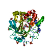
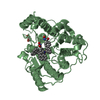
| Title | Crystal Structure of G303A HDAC8 in complex with M344 | ||||||
 Components Components | Histone deacetylase 8 | ||||||
 Keywords Keywords | HYDROLASE / Zinc Histone Deacetylase | ||||||
| Function / homology |  Function and homology information Function and homology informationhistone decrotonylase activity / histone deacetylase activity, hydrolytic mechanism / histone deacetylase / histone deacetylase activity / protein lysine deacetylase activity / Hydrolases; Acting on carbon-nitrogen bonds, other than peptide bonds; In linear amides / regulation of telomere maintenance / mitotic sister chromatid cohesion / Notch-HLH transcription pathway / nuclear chromosome ...histone decrotonylase activity / histone deacetylase activity, hydrolytic mechanism / histone deacetylase / histone deacetylase activity / protein lysine deacetylase activity / Hydrolases; Acting on carbon-nitrogen bonds, other than peptide bonds; In linear amides / regulation of telomere maintenance / mitotic sister chromatid cohesion / Notch-HLH transcription pathway / nuclear chromosome / histone deacetylase complex / negative regulation of protein ubiquitination / Hsp70 protein binding / Resolution of Sister Chromatid Cohesion / HDACs deacetylate histones / regulation of protein stability / Hsp90 protein binding / NOTCH1 Intracellular Domain Regulates Transcription / Constitutive Signaling by NOTCH1 PEST Domain Mutants / Constitutive Signaling by NOTCH1 HD+PEST Domain Mutants / Separation of Sister Chromatids / heterochromatin formation / chromatin organization / DNA-binding transcription factor binding / negative regulation of transcription by RNA polymerase II / nucleoplasm / metal ion binding / nucleus / cytoplasm Similarity search - Function | ||||||
| Biological species |  Homo sapiens (human) Homo sapiens (human) | ||||||
| Method |  X-RAY DIFFRACTION / SYNCHROTRON / MOLECULAR REPLACEMENT / Resolution: 2.407 Å X-RAY DIFFRACTION / SYNCHROTRON / MOLECULAR REPLACEMENT / Resolution: 2.407 Å | ||||||
 Authors Authors | Porter, N.J. / Christianson, D.W. | ||||||
| Funding support |  United States, 1items United States, 1items
| ||||||
 Citation Citation | Journal: Biochemistry / Year: 2016 Title: Structural and Functional Influence of the Glycine-Rich Loop G302GGGY on the Catalytic Tyrosine of Histone Deacetylase 8. Authors: Porter, N.J. / Christianson, N.H. / Decroos, C. / Christianson, D.W. | ||||||
| History |
|
- Structure visualization
| Structure viewer | Molecule: MolmilJmol/JSmol |
|---|
- Downloads & links
-Download
| PDBx/mmCIF format | 5tht.cif.gz | 296.1 KB | Display | PDBx/mmCIF format |
|---|---|---|---|---|
| PDB format | pdb5tht.ent.gz | 237.7 KB | Display | PDB format |
| PDBx/mmJSON format | 5tht.json.gz | Tree view | PDBx/mmJSON format | |
| Others |  Other downloads Other downloads |
-Validation report
| Arichive directory | https://data.pdbj.org/pub/pdb/validation_reports/th/5thtftp://data.pdbj.org/pub/pdb/validation_reports/th/5tht | HTTPS FTP |
|---|
-Related structure data
| Related structure data |  5thsC  5thuC  5thvC  3ewfS C: citing same article ( S: Starting model for refinement |
|---|---|
| Similar structure data |


-Links
 PDBj
PDBj


- Assembly
| Deposited unit | 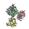
| ||||||||
|---|---|---|---|---|---|---|---|---|---|
| 1 | 
| ||||||||
| 2 | 
| ||||||||
| 3 | 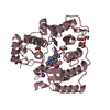
| ||||||||
| 4 | 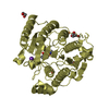
| ||||||||
| Unit cell |
|
-Components
-Protein , 1 types, 4 molecules ABCD
| #1: Protein | Mass: 43246.020 Da / Num. of mol.: 4 / Mutation: G303A Source method: isolated from a genetically manipulated source Source: (gene. exp.) Homo sapiens (human) / Gene: HDAC8, HDACL1, CDA07 / Plasmid: pET20b / Production host:  |
|---|
-Non-polymers , 5 types, 253 molecules 


| #2: Chemical | ChemComp-B3N /  Mass: 307.388 Da / Num. of mol.: 4 / Source method: obtained synthetically / Formula: C16H25N3O3 Mass: 307.388 Da / Num. of mol.: 4 / Source method: obtained synthetically / Formula: C16H25N3O3#3: Chemical | ChemComp-ZN /  Mass: 65.409 Da / Num. of mol.: 4 / Source method: obtained synthetically / Formula: Zn Mass: 65.409 Da / Num. of mol.: 4 / Source method: obtained synthetically / Formula: Zn#4: Chemical | ChemComp-K /  Mass: 39.098 Da / Num. of mol.: 8 / Source method: obtained synthetically / Formula: K Mass: 39.098 Da / Num. of mol.: 8 / Source method: obtained synthetically / Formula: K#5: Chemical | ChemComp-EDO /  Mass: 62.068 Da / Num. of mol.: 27 / Source method: obtained synthetically / Formula: C2H6O2 Mass: 62.068 Da / Num. of mol.: 27 / Source method: obtained synthetically / Formula: C2H6O2#6: Water | ChemComp-HOH / | Mass: 18.015 Da / Num. of mol.: 210 / Source method: isolated from a natural source / Formula: H2O |
|---|
-Experimental details
-Experiment
| Experiment | Method: X-RAY DIFFRACTION / Number of used crystals: 1 |
|---|
- Sample preparation
| Crystal | Density Matthews: 2.36 Å3/Da / Density % sol: 47.89 % |
|---|---|
| Crystal grow | Temperature: 294 K / Method: vapor diffusion, sitting drop / Details: 100 mM HEPES (pH 7.5), 22% w/v PEG 6000, 4 mM TCEP |
-Data collection
| Diffraction | Mean temperature: 100 K |
|---|---|
| Diffraction source | Source: SYNCHROTRON / Site: APS / Beamline: 24-ID-E / Wavelength: 0.97923 Å |
| Detector | Type: ADSC QUANTUM 315 / Detector: CCD / Date: Nov 9, 2015 |
| Radiation | Protocol: SINGLE WAVELENGTH / Monochromatic (M) / Laue (L): M / Scattering type: x-ray |
| Radiation wavelength | Wavelength: 0.97923 Å / Relative weight: 1 |
| Reflection | Resolution: 2.407→97.66 Å / Num. obs: 62588 / % possible obs: 99.9 % / Redundancy: 3.5 % / CC1/2: 0.996 / Rmerge(I) obs: 0.068 / Rsym value: 0.066 / Net I/σ(I): 12.5 |
- Processing
| Software |
| ||||||||||||||||||||||||||||||||||||||||||||||||||||||||||||||||||||||||||||||||||||||||||||||||||||||||||||||||||||||||||||||||||||||||||||||||||||||||||
|---|---|---|---|---|---|---|---|---|---|---|---|---|---|---|---|---|---|---|---|---|---|---|---|---|---|---|---|---|---|---|---|---|---|---|---|---|---|---|---|---|---|---|---|---|---|---|---|---|---|---|---|---|---|---|---|---|---|---|---|---|---|---|---|---|---|---|---|---|---|---|---|---|---|---|---|---|---|---|---|---|---|---|---|---|---|---|---|---|---|---|---|---|---|---|---|---|---|---|---|---|---|---|---|---|---|---|---|---|---|---|---|---|---|---|---|---|---|---|---|---|---|---|---|---|---|---|---|---|---|---|---|---|---|---|---|---|---|---|---|---|---|---|---|---|---|---|---|---|---|---|---|---|---|---|---|
| Refinement | Method to determine structure: MOLECULAR REPLACEMENT Starting model: 3EWF Resolution: 2.407→97.656 Å / SU ML: 0.23 / Cross valid method: FREE R-VALUE / σ(F): 1.35 / Phase error: 20.06
| ||||||||||||||||||||||||||||||||||||||||||||||||||||||||||||||||||||||||||||||||||||||||||||||||||||||||||||||||||||||||||||||||||||||||||||||||||||||||||
| Solvent computation | Shrinkage radii: 0.9 Å / VDW probe radii: 1.11 Å | ||||||||||||||||||||||||||||||||||||||||||||||||||||||||||||||||||||||||||||||||||||||||||||||||||||||||||||||||||||||||||||||||||||||||||||||||||||||||||
| Refinement step | Cycle: LAST / Resolution: 2.407→97.656 Å
| ||||||||||||||||||||||||||||||||||||||||||||||||||||||||||||||||||||||||||||||||||||||||||||||||||||||||||||||||||||||||||||||||||||||||||||||||||||||||||
| Refine LS restraints |
| ||||||||||||||||||||||||||||||||||||||||||||||||||||||||||||||||||||||||||||||||||||||||||||||||||||||||||||||||||||||||||||||||||||||||||||||||||||||||||
| LS refinement shell |
|
