 Movie
Movie Controller

[English] 日本語
 Yorodumi
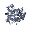
- PDB-7jvw: Crystal structure of human histone deacetylase 8 (HDAC8) G320R mu... -
+ Open data

- Basic information
| Entry | Database: PDB / ID: 7jvw | ||||||
|---|---|---|---|---|---|---|---|
| Title | Crystal structure of human histone deacetylase 8 (HDAC8) G320R mutation complexed with M344 | ||||||
 Components Components | Histone deacetylase 8 | ||||||
 Keywords Keywords | HYDROLASE / Histone deacetylase / Cornelia de Lange Syndrome (CdLS) | ||||||
| Function / homology |  Function and homology information Function and homology informationhistone decrotonylase activity / histone deacetylase activity, hydrolytic mechanism / histone deacetylase / histone deacetylase activity / protein lysine deacetylase activity / Hydrolases; Acting on carbon-nitrogen bonds, other than peptide bonds; In linear amides / regulation of telomere maintenance / mitotic sister chromatid cohesion / Notch-HLH transcription pathway / nuclear chromosome ...histone decrotonylase activity / histone deacetylase activity, hydrolytic mechanism / histone deacetylase / histone deacetylase activity / protein lysine deacetylase activity / Hydrolases; Acting on carbon-nitrogen bonds, other than peptide bonds; In linear amides / regulation of telomere maintenance / mitotic sister chromatid cohesion / Notch-HLH transcription pathway / nuclear chromosome / histone deacetylase complex / negative regulation of protein ubiquitination / Hsp70 protein binding / Resolution of Sister Chromatid Cohesion / HDACs deacetylate histones / regulation of protein stability / Hsp90 protein binding / NOTCH1 Intracellular Domain Regulates Transcription / Constitutive Signaling by NOTCH1 PEST Domain Mutants / Constitutive Signaling by NOTCH1 HD+PEST Domain Mutants / Separation of Sister Chromatids / heterochromatin formation / chromatin organization / DNA-binding transcription factor binding / negative regulation of transcription by RNA polymerase II / nucleoplasm / metal ion binding / nucleus / cytoplasm Similarity search - Function | ||||||
| Biological species |  Homo sapiens (human) Homo sapiens (human) | ||||||
| Method |  X-RAY DIFFRACTION / SYNCHROTRON / MOLECULAR REPLACEMENT / Resolution: 2.40301779656 Å X-RAY DIFFRACTION / SYNCHROTRON / MOLECULAR REPLACEMENT / Resolution: 2.40301779656 Å | ||||||
 Authors Authors | Osko, J.D. / Christianson, D.W. / Decroos, C. / Porter, N.J. / Lee, M. | ||||||
| Funding support |  United States, 1items United States, 1items
| ||||||
 Citation Citation | Journal: J.Struct.Biol. / Year: 2020 Title: Structural analysis of histone deacetylase 8 mutants associated with Cornelia de Lange Syndrome spectrum disorders. Authors: Osko, J.D. / Porter, N.J. / Decroos, C. / Lee, M.S. / Watson, P.R. / Raible, S.E. / Krantz, I.D. / Deardorff, M.A. / Christianson, D.W. | ||||||
| History |
|
- Structure visualization
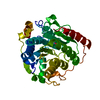
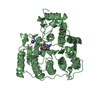
| Structure viewer | Molecule: MolmilJmol/JSmol |
|---|
- Downloads & links
-Download
| PDBx/mmCIF format | 7jvw.cif.gz | 187.1 KB | Display | PDBx/mmCIF format |
|---|---|---|---|---|
| PDB format | pdb7jvw.ent.gz | 117.4 KB | Display | PDB format |
| PDBx/mmJSON format | 7jvw.json.gz | Tree view | PDBx/mmJSON format | |
| Others |  Other downloads Other downloads |
-Validation report
| Arichive directory | https://data.pdbj.org/pub/pdb/validation_reports/jv/7jvwftp://data.pdbj.org/pub/pdb/validation_reports/jv/7jvw | HTTPS FTP |
|---|
-Related structure data
| Related structure data |  7jvuC  7jvvC  3ewfS S: Starting model for refinement C: citing same article ( |
|---|---|
| Similar structure data |


-Links
 PDBj
PDBj


- Assembly
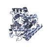
| Deposited unit | 
| ||||||||||||
|---|---|---|---|---|---|---|---|---|---|---|---|---|---|
| 1 | 
| ||||||||||||
| 2 | 
| ||||||||||||
| Unit cell |
|
-Components
| #1: Protein | Mass: 43332.137 Da / Num. of mol.: 2 / Mutation: G320R Source method: isolated from a genetically manipulated source Source: (gene. exp.) Homo sapiens (human) / Gene: HDAC8, HDACL1, CDA07 / Production host:  #2: Chemical | 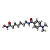  Mass: 307.388 Da / Num. of mol.: 2 / Source method: obtained synthetically / Formula: C16H25N3O3 Mass: 307.388 Da / Num. of mol.: 2 / Source method: obtained synthetically / Formula: C16H25N3O3#3: Chemical |   Mass: 65.409 Da / Num. of mol.: 2 / Source method: obtained synthetically / Formula: Zn Mass: 65.409 Da / Num. of mol.: 2 / Source method: obtained synthetically / Formula: Zn#4: Chemical | ChemComp-K /   Mass: 39.098 Da / Num. of mol.: 4 / Source method: obtained synthetically / Formula: K Mass: 39.098 Da / Num. of mol.: 4 / Source method: obtained synthetically / Formula: K#5: Water | ChemComp-HOH / |  Mass: 18.015 Da / Num. of mol.: 143 / Source method: isolated from a natural source / Formula: H2O Mass: 18.015 Da / Num. of mol.: 143 / Source method: isolated from a natural source / Formula: H2OHas ligand of interest | N | |
|---|
-Experimental details
-Experiment
| Experiment | Method: X-RAY DIFFRACTION / Number of used crystals: 1 |
|---|
- Sample preparation
| Crystal | Density Matthews: 2.36 Å3/Da / Density % sol: 47.93 % |
|---|---|
| Crystal grow | Temperature: 294 K / Method: vapor diffusion, sitting drop / pH: 5.3 / Details: 0.1 M MES, pH 5.3, 3% PEG1500, 4 mM TCEP |
-Data collection
| Diffraction | Mean temperature: 100 K / Serial crystal experiment: N |
|---|---|
| Diffraction source | Source: SYNCHROTRON / Site: SSRL / Beamline: BL14-1 / Wavelength: 1 Å |
| Detector | Type: MARMOSAIC 325 mm CCD / Detector: CCD / Date: Dec 9, 2015 |
| Radiation | Monochromator: double crystal Si(111) / Protocol: SINGLE WAVELENGTH / Monochromatic (M) / Laue (L): M / Scattering type: x-ray |
| Radiation wavelength | Wavelength: 1 Å / Relative weight: 1 |
| Reflection | Resolution: 2.4→82.75 Å / Num. obs: 31564 / % possible obs: 97.4 % / Redundancy: 3.8 % / Biso Wilson estimate: 34.6910203953 Å2 / CC1/2: 0.981 / Rmerge(I) obs: 0.148 / Rpim(I) all: 0.09 / Net I/σ(I): 8.7 |
| Reflection shell | Resolution: 2.4→2.49 Å / Redundancy: 3.8 % / Rmerge(I) obs: 0.961 / Mean I/σ(I) obs: 1.7 / Num. unique obs: 30670 / CC1/2: 0.578 / Rpim(I) all: 0.571 / % possible all: 96 |
- Processing
| Software |
| ||||||||||||||||||||||||||||||||||||||||||||||||||||||||||||||||||||||||||||||||||||
|---|---|---|---|---|---|---|---|---|---|---|---|---|---|---|---|---|---|---|---|---|---|---|---|---|---|---|---|---|---|---|---|---|---|---|---|---|---|---|---|---|---|---|---|---|---|---|---|---|---|---|---|---|---|---|---|---|---|---|---|---|---|---|---|---|---|---|---|---|---|---|---|---|---|---|---|---|---|---|---|---|---|---|---|---|---|
| Refinement | Method to determine structure: MOLECULAR REPLACEMENT Starting model: PDB entry 3EWF Resolution: 2.40301779656→48.9900201109 Å / SU ML: 0.381622707936 / Cross valid method: FREE R-VALUE / σ(F): 1.33674203891 / Phase error: 29.5397908894 Stereochemistry target values: GeoStd + Monomer Library + CDL v1.2
| ||||||||||||||||||||||||||||||||||||||||||||||||||||||||||||||||||||||||||||||||||||
| Solvent computation | Shrinkage radii: 0.9 Å / VDW probe radii: 1.11 Å / Solvent model: FLAT BULK SOLVENT MODEL | ||||||||||||||||||||||||||||||||||||||||||||||||||||||||||||||||||||||||||||||||||||
| Displacement parameters | Biso mean: 40.6687983774 Å2 | ||||||||||||||||||||||||||||||||||||||||||||||||||||||||||||||||||||||||||||||||||||
| Refinement step | Cycle: LAST / Resolution: 2.40301779656→48.9900201109 Å
| ||||||||||||||||||||||||||||||||||||||||||||||||||||||||||||||||||||||||||||||||||||
| Refine LS restraints |
| ||||||||||||||||||||||||||||||||||||||||||||||||||||||||||||||||||||||||||||||||||||
| LS refinement shell |
|
