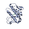
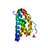
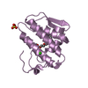
GolgiresidentproteinGCP60 / Acyl-CoA-binding domain-containing protein 3 / Golgi complex-associated protein 1 / GOCAP1 / Golgi ...Acyl-CoA-binding domain-containing protein 3 / Golgi complex-associated protein 1 / GOCAP1 / Golgi phosphoprotein 1 / GOLPH1 / PBR- and PKA-associated protein 7 / Peripheral benzodiazepine receptor-associated protein PAP7
Mass: 19214.143 Da / Num. of mol.: 1 / Fragment: GOLD domain, UNP residues 367-528 Source method: isolated from a genetically manipulated source Source: (gene. exp.) Homo sapiens (human) / Gene: ACBD3, GCP60, GOCAP1, GOLPH1 / Plasmid: pOPTH / Production host: Escherichia coli (E. coli) / Strain (production host): BL21(DE3) / References: UniProt: Q9H3P7
-
Experimental details
-
Experiment
Experiment
Method: X-RAY DIFFRACTION / Number of used crystals: 1
-
Sample preparation
Crystal
Density Matthews: 3.24 Å3/Da / Density % sol: 62.08 % Preparation: The low data completeness is due to the data being severely anisotropic, and the data was processed using the diffraction anisotropy server (https://services.mbi.ucla.edu/anisoscale/). ...Preparation: The low data completeness is due to the data being severely anisotropic, and the data was processed using the diffraction anisotropy server (https://services.mbi.ucla.edu/anisoscale/). Data was truncated and scaled with dimensions a*b*c*- 3.2,3.1,2.5. This is the reason for the low completeness in the resolution shells from 3.1 to 2.5. This processing was the only way to generate maps of high enough quality for model building
Crystal grow
Temperature: 289 K / Method: vapor diffusion, hanging drop / pH: 7.5 Details: 13% PEG 4000, 200 mM Ammonium Sulfate, 100 mM HEPES, 10% 2-propanol, 10% glycerol
In the structure databanks used in Yorodumi, some data are registered as the other names, "COVID-19 virus" and "2019-nCoV". Here are the details of the virus and the list of structure data.
Jan 31, 2019. EMDB accession codes are about to change! (news from PDBe EMDB page)
EMDB accession codes are about to change! (news from PDBe EMDB page)
The allocation of 4 digits for EMDB accession codes will soon come to an end. Whilst these codes will remain in use, new EMDB accession codes will include an additional digit and will expand incrementally as the available range of codes is exhausted. The current 4-digit format prefixed with “EMD-” (i.e. EMD-XXXX) will advance to a 5-digit format (i.e. EMD-XXXXX), and so on. It is currently estimated that the 4-digit codes will be depleted around Spring 2019, at which point the 5-digit format will come into force.
The EM Navigator/Yorodumi systems omit the EMD- prefix.
Related info.:Q: What is EMD? / ID/Accession-code notation in Yorodumi/EM Navigator
Yorodumi is a browser for structure data from EMDB, PDB, SASBDB, etc.
This page is also the successor to EM Navigator detail page, and also detail information page/front-end page for Omokage search.
The word "yorodu" (or yorozu) is an old Japanese word meaning "ten thousand". "mi" (miru) is to see.
Related info.:EMDB / PDB / SASBDB / Comparison of 3 databanks / Yorodumi Search / Aug 31, 2016. New EM Navigator & Yorodumi / Yorodumi Papers / Jmol/JSmol / Function and homology information / Changes in new EM Navigator and Yorodumi
 Movie
Movie Controller


 Open data
 Basic information
 Components
 Keywords
 Function and homology information
 Homo sapiens (human)
 X-RAY DIFFRACTION /
  Authors
 Canada, 1items
  Citation
 Structure visualization
 Downloads & links
 Other downloads


 PDBj
PDBj

 Assembly
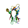

 Sample preparation
 Processing