 Movie
Movie Controller


+ Open data

- Basic information
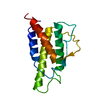
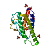
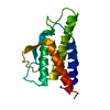
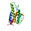
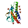
| Entry | Database: PDB / ID: 1hzi | ||||||
|---|---|---|---|---|---|---|---|
| Title | INTERLEUKIN-4 MUTANT E9A | ||||||
 Components Components | INTERLEUKIN-4 | ||||||
 Keywords Keywords | CYTOKINE / IL-4 / 4-HELIX-BUNDLE | ||||||
| Function / homology |  Function and homology information Function and homology informationinterleukin-4 receptor binding / positive regulation of isotype switching to IgE isotypes / positive regulation of cellular respiration / negative regulation of complement-dependent cytotoxicity / Interleukin-18 signaling / regulation of isotype switching / negative regulation of neuroinflammatory response / negative regulation of epithelial cell migration / positive regulation of T-helper 2 cell cytokine production / dendritic cell differentiation ...interleukin-4 receptor binding / positive regulation of isotype switching to IgE isotypes / positive regulation of cellular respiration / negative regulation of complement-dependent cytotoxicity / Interleukin-18 signaling / regulation of isotype switching / negative regulation of neuroinflammatory response / negative regulation of epithelial cell migration / positive regulation of T-helper 2 cell cytokine production / dendritic cell differentiation / interleukin-4-mediated signaling pathway / neuroinflammatory response / positive regulation of isotype switching to IgG isotypes / positive regulation of interleukin-13 production / macrophage activation / positive regulation of amyloid-beta clearance / myeloid dendritic cell differentiation / positive regulation of MHC class II biosynthetic process / type 2 immune response / negative regulation of cellular response to transforming growth factor beta stimulus / positive regulation of T cell differentiation / negative regulation of osteoclast differentiation / positive regulation of ATP biosynthetic process / positive regulation of macroautophagy / positive regulation of interleukin-10 production / negative regulation of tumor necrosis factor production / regulation of immune response / negative regulation of endothelial cell apoptotic process / cell surface receptor signaling pathway via JAK-STAT / cholesterol metabolic process / positive regulation of B cell proliferation / positive regulation of T cell proliferation / B cell differentiation / cytokine activity / T cell activation / growth factor activity / positive regulation of receptor-mediated endocytosis / negative regulation of inflammatory response / positive regulation of cold-induced thermogenesis / Interleukin-4 and Interleukin-13 signaling / immune response / positive regulation of cell migration / negative regulation of DNA-templated transcription / positive regulation of cell population proliferation / positive regulation of gene expression / negative regulation of apoptotic process / positive regulation of DNA-templated transcription / negative regulation of transcription by RNA polymerase II / positive regulation of transcription by RNA polymerase II / : / extracellular region Similarity search - Function | ||||||
| Biological species |  Homo sapiens (human) Homo sapiens (human) | ||||||
| Method |  X-RAY DIFFRACTION / MOLECULAR REPLACEMENT / Resolution: 2.05 Å X-RAY DIFFRACTION / MOLECULAR REPLACEMENT / Resolution: 2.05 Å | ||||||
 Authors Authors | Hulsmeyer, M. / Scheufler, C. / Dreyer, M.K. | ||||||
 Citation Citation | Journal: Acta Crystallogr.,Sect.D / Year: 2001 Title: Structure of interleukin 4 mutant E9A suggests polar steering in receptor-complex formation. Authors: Hulsmeyer, M. / Scheufler, C. / Dreyer, M.K. #1: Journal: Proc.Natl.Acad.Sci.USA / Year: 1997Title: A MIXED-CHARGE PAIR IN HUMAN INTERLEUKIN 4 DOMINATES HIGH-AFFINITY INTERACTION WITH THE RECEPTOR A CHAIN Authors: Wang, Y. / Shen, B.-J. / Sebald, W. #2: Journal: Cell(Cambridge,Mass.) / Year: 1999Title: Crystal Structure of the Interleukin-4/Receptor a chain complex reveals a mosaic binding interface Authors: Hage, T. / Sebald, W. / Reinemer, P. | ||||||
| History |
|
- Structure visualization
| Structure viewer | Molecule: MolmilJmol/JSmol |
|---|
- Downloads & links
-Download
| PDBx/mmCIF format | 1hzi.cif.gz | 43.3 KB | Display | PDBx/mmCIF format |
|---|---|---|---|---|
| PDB format | pdb1hzi.ent.gz | 29.8 KB | Display | PDB format |
| PDBx/mmJSON format | 1hzi.json.gz | Tree view | PDBx/mmJSON format | |
| Others |  Other downloads Other downloads |
-Validation report
| Arichive directory | https://data.pdbj.org/pub/pdb/validation_reports/hz/1hziftp://data.pdbj.org/pub/pdb/validation_reports/hz/1hzi | HTTPS FTP |
|---|
-Related structure data
| Related structure data |  1rcbS S: Starting model for refinement |
|---|---|
| Similar structure data |


-Links
 PDBj
PDBj


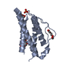
- Assembly
| Deposited unit | 
| ||||||||
|---|---|---|---|---|---|---|---|---|---|
| 1 |
| ||||||||
| Unit cell |
| ||||||||
| Details | The biological assembly is a monomer. |
-Components
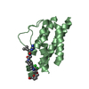
| #1: Protein | Mass: 14931.213 Da / Num. of mol.: 1 / Mutation: E9A Source method: isolated from a genetically manipulated source Source: (gene. exp.) Homo sapiens (human) / Production host:  | ||||
|---|---|---|---|---|---|
| #2: Chemical | ChemComp-SO4 /   Mass: 96.063 Da / Num. of mol.: 5 / Source method: obtained synthetically / Formula: SO4 Mass: 96.063 Da / Num. of mol.: 5 / Source method: obtained synthetically / Formula: SO4#3: Water | ChemComp-HOH / |  Mass: 18.015 Da / Num. of mol.: 145 / Source method: isolated from a natural source / Formula: H2O Mass: 18.015 Da / Num. of mol.: 145 / Source method: isolated from a natural source / Formula: H2OHas protein modification | Y | |
-Experimental details
-Experiment
| Experiment | Method: X-RAY DIFFRACTION / Number of used crystals: 1 |
|---|
- Sample preparation
| Crystal | Density Matthews: 3.15 Å3/Da / Density % sol: 60 % | ||||||||||||||||||||
|---|---|---|---|---|---|---|---|---|---|---|---|---|---|---|---|---|---|---|---|---|---|
| Crystal grow | Temperature: 293 K / Method: vapor diffusion, hanging drop / pH: 5.4 Details: ammonium sulfate, sodium acetate, pH 5.4, VAPOR DIFFUSION, HANGING DROP, temperature 293K | ||||||||||||||||||||
| Crystal grow | *PLUS | ||||||||||||||||||||
| Components of the solutions | *PLUS
|
-Data collection
| Diffraction | Mean temperature: 100 K |
|---|---|
| Diffraction source | Source: ROTATING ANODE / Type: RIGAKU RU200 / Wavelength: 1.5418 Å |
| Detector | Type: MARRESEARCH / Detector: IMAGE PLATE / Date: Jun 11, 1998 |
| Radiation | Monochromator: GRAPHITE / Protocol: SINGLE WAVELENGTH / Monochromatic (M) / Laue (L): M / Scattering type: x-ray |
| Radiation wavelength | Wavelength: 1.5418 Å / Relative weight: 1 |
| Reflection | Resolution: 2.05→20 Å / Num. all: 12421 / Num. obs: 12421 / % possible obs: 99.1 % / Observed criterion σ(F): 0 / Observed criterion σ(I): 0 / Redundancy: 5 % / Biso Wilson estimate: 21.5 Å2 / Rmerge(I) obs: 0.037 / Net I/σ(I): 34.8 |
| Reflection shell | Resolution: 2.05→2.1 Å / Redundancy: 4.6 % / Rmerge(I) obs: 0.168 / Mean I/σ(I) obs: 7.7 / Num. unique all: 795 / % possible all: 98.8 |
| Reflection | *PLUS Lowest resolution: 20 Å / % possible obs: 99.2 % / Num. measured all: 61298 / Rmerge(I) obs: 0.034 |
| Reflection shell | *PLUS % possible obs: 98.8 % / Num. unique obs: 795 / Num. measured obs: 4531 |
- Processing
| Software |
| ||||||||||||||||||||||||||||||||||||||||
|---|---|---|---|---|---|---|---|---|---|---|---|---|---|---|---|---|---|---|---|---|---|---|---|---|---|---|---|---|---|---|---|---|---|---|---|---|---|---|---|---|---|
| Refinement | Method to determine structure: MOLECULAR REPLACEMENT Starting model: PDB ENTRY 1RCB Resolution: 2.05→19.88 Å / Rfactor Rfree error: 0.008 / Data cutoff high absF: 1148676.14 / Data cutoff low absF: 0 / Isotropic thermal model: RESTRAINED / Cross valid method: THROUGHOUT / σ(F): 0 / σ(I): 0 / Stereochemistry target values: Engh & Huber
| ||||||||||||||||||||||||||||||||||||||||
| Solvent computation | Solvent model: FLAT MODEL / Bsol: 55.42 Å2 / ksol: 0.377 e/Å3 | ||||||||||||||||||||||||||||||||||||||||
| Displacement parameters | Biso mean: 32.5 Å2
| ||||||||||||||||||||||||||||||||||||||||
| Refine analyze |
| ||||||||||||||||||||||||||||||||||||||||
| Refinement step | Cycle: LAST / Resolution: 2.05→19.88 Å
| ||||||||||||||||||||||||||||||||||||||||
| Refine LS restraints |
| ||||||||||||||||||||||||||||||||||||||||
| LS refinement shell | Resolution: 2.05→2.18 Å / Rfactor Rfree error: 0.023 / Total num. of bins used: 6
| ||||||||||||||||||||||||||||||||||||||||
| Xplor file |
| ||||||||||||||||||||||||||||||||||||||||
| Software | *PLUS Name: CNS / Classification: refinement | ||||||||||||||||||||||||||||||||||||||||
| Refinement | *PLUS σ(F): 0 / % reflection Rfree: 7.1 % | ||||||||||||||||||||||||||||||||||||||||
| Solvent computation | *PLUS | ||||||||||||||||||||||||||||||||||||||||
| Displacement parameters | *PLUS Biso mean: 32.5 Å2 | ||||||||||||||||||||||||||||||||||||||||
| Refine LS restraints | *PLUS
| ||||||||||||||||||||||||||||||||||||||||
| LS refinement shell | *PLUS Rfactor Rfree: 0.285 / % reflection Rfree: 7.7 % / Rfactor Rwork: 0.252 |
