 Movie
Movie Controller

[English] 日本語
 Yorodumi
- PDB-2i0u: Crystal structures of phospholipases A2 from Vipera nikolskii ven... -
+ Open data

- Basic information
| Entry | Database: PDB / ID: 2i0u | ||||||
|---|---|---|---|---|---|---|---|
| Title | Crystal structures of phospholipases A2 from Vipera nikolskii venom revealing Triton X-100 bound in hydrophobic channel | ||||||
 Components Components | Basic subunit of heterodimer phospholipase A2 | ||||||
 Keywords Keywords | HYDROLASE / alpha-beta-alpha | ||||||
| Function / homology |  Function and homology information Function and homology informationphospholipase A2 / : / arachidonate secretion / lipid catabolic process / negative regulation of T cell proliferation / phospholipid metabolic process / phospholipid binding / toxin activity / calcium ion binding / extracellular region Similarity search - Function | ||||||
| Biological species |  Vipera nikolskii (snake) Vipera nikolskii (snake) | ||||||
| Method |  X-RAY DIFFRACTION / SYNCHROTRON / MOLECULAR REPLACEMENT / Resolution: 2.2 Å X-RAY DIFFRACTION / SYNCHROTRON / MOLECULAR REPLACEMENT / Resolution: 2.2 Å | ||||||
 Authors Authors | Gao, W. / Bi, R.C. | ||||||
 Citation Citation | Journal: To be Published Title: Crystal structures of phospholipases A2 from Vipera nikolskii venom revealing Triton X-100 bound in hydrophobic channel Authors: Gao, W. / Bi, R.C. | ||||||
| History |
|
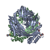
- Structure visualization
| Structure viewer | Molecule: MolmilJmol/JSmol |
|---|
- Downloads & links
-Download
| PDBx/mmCIF format | 2i0u.cif.gz | 68.4 KB | Display | PDBx/mmCIF format |
|---|---|---|---|---|
| PDB format | pdb2i0u.ent.gz | 50.1 KB | Display | PDB format |
| PDBx/mmJSON format | 2i0u.json.gz | Tree view | PDBx/mmJSON format | |
| Others |  Other downloads Other downloads |
-Validation report
| Arichive directory | https://data.pdbj.org/pub/pdb/validation_reports/i0/2i0uftp://data.pdbj.org/pub/pdb/validation_reports/i0/2i0u | HTTPS FTP |
|---|
-Related structure data
| Similar structure data |
|---|
-Links
 PDBj
PDBj

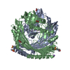
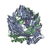
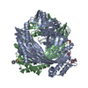
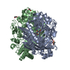
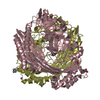
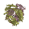
- Assembly
| Deposited unit | 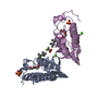
| |||||||||
|---|---|---|---|---|---|---|---|---|---|---|
| 1 | 
| |||||||||
| 2 | 
| |||||||||
| 3 | 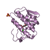
| |||||||||
| Unit cell |
| |||||||||
| Components on special symmetry positions |
| |||||||||
| Details | The second part of the biological assembly is generated by the three fold axis: x+2/3, y+1/3, z. |
-Components
-Protein , 1 types, 2 molecules EA
| #1: Protein | Mass: 13829.771 Da / Num. of mol.: 2 / Source method: isolated from a natural source / Source: (natural) Vipera nikolskii (snake) / Secretion: venom / References: UniProt: Q1RP79, phospholipase A2 |
|---|
-Non-polymers , 5 types, 215 molecules 


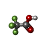

| #2: Chemical |  Mass: 40.078 Da / Num. of mol.: 2 / Source method: obtained synthetically / Formula: Ca Mass: 40.078 Da / Num. of mol.: 2 / Source method: obtained synthetically / Formula: Ca#3: Chemical |  Mass: 96.063 Da / Num. of mol.: 2 / Source method: obtained synthetically / Formula: SO4 Mass: 96.063 Da / Num. of mol.: 2 / Source method: obtained synthetically / Formula: SO4#4: Chemical |  Mass: 352.508 Da / Num. of mol.: 2 / Source method: obtained synthetically / Formula: C21H36O4 / Comment: detergent*YM Mass: 352.508 Da / Num. of mol.: 2 / Source method: obtained synthetically / Formula: C21H36O4 / Comment: detergent*YM#5: Chemical | ChemComp-TFA / |  Mass: 114.023 Da / Num. of mol.: 1 / Source method: obtained synthetically / Formula: C2HF3O2 Mass: 114.023 Da / Num. of mol.: 1 / Source method: obtained synthetically / Formula: C2HF3O2#6: Water | ChemComp-HOH / | Mass: 18.015 Da / Num. of mol.: 208 / Source method: isolated from a natural source / Formula: H2O |
|---|
-Details
| Has protein modification | Y |
|---|
-Experimental details
-Experiment
| Experiment | Method: X-RAY DIFFRACTION / Number of used crystals: 1 |
|---|
- Sample preparation
| Crystal | Density Matthews: 3.08 Å3/Da / Density % sol: 60.07 % |
|---|---|
| Crystal grow | Temperature: 291 K / Method: vapor diffusion, hanging drop / pH: 6.5 Details: 15% PEG 4K, 0.2M lithium sulphate, 0.1M sodium cacodylate pH 6.5, 0.1% sodium azide, VAPOR DIFFUSION, HANGING DROP, temperature 291K |
-Data collection
| Diffraction | Mean temperature: 298 K |
|---|---|
| Diffraction source | Source: SYNCHROTRON / Site: BSRF  / Beamline: 3W1A / Wavelength: 1 Å / Beamline: 3W1A / Wavelength: 1 Å |
| Detector | Type: MAR CCD 165 mm / Detector: CCD |
| Radiation | Protocol: SINGLE WAVELENGTH / Monochromatic (M) / Laue (L): M / Scattering type: x-ray |
| Radiation wavelength | Wavelength: 1 Å / Relative weight: 1 |
| Reflection | Resolution: 2.2→29 Å / Num. obs: 17760 / % possible obs: 90.2 % / Observed criterion σ(F): 0 / Observed criterion σ(I): 0 |
| Reflection shell | Resolution: 2.2→2.26 Å |
- Processing
| Software |
| ||||||||||||||||||
|---|---|---|---|---|---|---|---|---|---|---|---|---|---|---|---|---|---|---|---|
| Refinement | Method to determine structure: MOLECULAR REPLACEMENT / Resolution: 2.2→29 Å / σ(F): 2
| ||||||||||||||||||
| Refinement step | Cycle: LAST / Resolution: 2.2→29 Å
| ||||||||||||||||||
| Refine LS restraints |
|
