 Movie
Movie Controller

[English] 日本語
 Yorodumi
- PDB-4jl0: Crystal structure of PcrH in complex with the chaperone binding r... -
+ Open data

- Basic information
| Entry | Database: PDB / ID: 4jl0 | ||||||
|---|---|---|---|---|---|---|---|
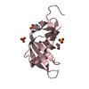
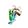
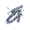
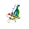
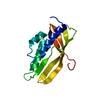
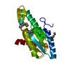
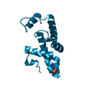
| Title | Crystal structure of PcrH in complex with the chaperone binding region of PopB | ||||||
 Components Components |
| ||||||
 Keywords Keywords | CHAPERONE / translocator / protein binding / type III secretion system / TPR / tetratricopeptide repeat | ||||||
| Function / homology |  Function and homology information Function and homology information | ||||||
| Biological species |   Pseudomonas aeruginosa (bacteria) Pseudomonas aeruginosa (bacteria) | ||||||
| Method |  X-RAY DIFFRACTION / SYNCHROTRON / MOLECULAR REPLACEMENT / molecular replacement / Resolution: 2.22 Å X-RAY DIFFRACTION / SYNCHROTRON / MOLECULAR REPLACEMENT / molecular replacement / Resolution: 2.22 Å | ||||||
 Authors Authors | Discola, K.F. / Forster, A. / Simorre, J.P. / Attree, I. / Dessen, A. / Job, V. | ||||||
 Citation Citation | Journal: J.Biol.Chem. / Year: 2014 Title: Membrane and Chaperone Recognition by the Major Translocator Protein PopB of the Type III Secretion System of Pseudomonas aeruginosa. Authors: Discola, K.F. / Forster, A. / Boulay, F. / Simorre, J.P. / Attree, I. / Dessen, A. / Job, V. | ||||||
| History |
|
- Structure visualization
| Structure viewer | Molecule: MolmilJmol/JSmol |
|---|
- Downloads & links
-Download
| PDBx/mmCIF format | 4jl0.cif.gz | 66.6 KB | Display | PDBx/mmCIF format |
|---|---|---|---|---|
| PDB format | pdb4jl0.ent.gz | 48 KB | Display | PDB format |
| PDBx/mmJSON format | 4jl0.json.gz | Tree view | PDBx/mmJSON format | |
| Others |  Other downloads Other downloads |
-Validation report
| Arichive directory | https://data.pdbj.org/pub/pdb/validation_reports/jl/4jl0ftp://data.pdbj.org/pub/pdb/validation_reports/jl/4jl0 | HTTPS FTP |
|---|
-Related structure data
| Related structure data | 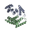 2xccS S: Starting model for refinement |
|---|---|
| Similar structure data |


-Links
 PDBj
PDBj
- Assembly
| Deposited unit | 
| ||||||||
|---|---|---|---|---|---|---|---|---|---|
| 1 | 
| ||||||||
| 2 | 
| ||||||||
| Unit cell |
| ||||||||
| Details | There are 2 biological units in the asymmetric unit |
-Components
| #1: Protein | Mass: 15486.300 Da / Num. of mol.: 2 / Fragment: UNP residues 22-161 Source method: isolated from a genetically manipulated source Source: (gene. exp.) Pseudomonas aeruginosa (bacteria) / Strain: CHA / Gene: pcrH / Plasmid: pETDUET1 / Production host: #2: Protein/peptide | Mass: 841.948 Da / Num. of mol.: 2 / Fragment: UNP residues 51-59 / Source method: obtained synthetically / Details: synthesized peptide / Source: (synth.) Pseudomonas aeruginosa (bacteria) / References: UniProt: Q840U9#3: Chemical | ChemComp-TRS / |   Mass: 122.143 Da / Num. of mol.: 1 / Source method: obtained synthetically / Formula: C4H12NO3 / Comment: pH buffer*YM Mass: 122.143 Da / Num. of mol.: 1 / Source method: obtained synthetically / Formula: C4H12NO3 / Comment: pH buffer*YM#4: Water | ChemComp-HOH / |  Mass: 18.015 Da / Num. of mol.: 113 / Source method: isolated from a natural source / Formula: H2O Mass: 18.015 Da / Num. of mol.: 113 / Source method: isolated from a natural source / Formula: H2O |
|---|
-Experimental details
-Experiment
| Experiment | Method: X-RAY DIFFRACTION / Number of used crystals: 1 |
|---|
- Sample preparation
| Crystal | Density Matthews: 2.5 Å3/Da / Density % sol: 50.87 % |
|---|---|
| Crystal grow | Temperature: 293 K / Method: vapor diffusion, hanging drop / pH: 8 Details: 30% PEG6000, 1.0M LiCl/ 0.1 M tris-HCl pH 8.0, VAPOR DIFFUSION, HANGING DROP, temperature 293K |
-Data collection
| Diffraction | Mean temperature: 100 K | ||||||||||||||||||||||||||||||||||||||||||||||||||||||||||||||||||||||
|---|---|---|---|---|---|---|---|---|---|---|---|---|---|---|---|---|---|---|---|---|---|---|---|---|---|---|---|---|---|---|---|---|---|---|---|---|---|---|---|---|---|---|---|---|---|---|---|---|---|---|---|---|---|---|---|---|---|---|---|---|---|---|---|---|---|---|---|---|---|---|---|
| Diffraction source | Source: SYNCHROTRON / Site: ESRF  / Beamline: BM14 / Wavelength: 0.97796 Å / Beamline: BM14 / Wavelength: 0.97796 Å | ||||||||||||||||||||||||||||||||||||||||||||||||||||||||||||||||||||||
| Detector | Type: MARMOSAIC 225 mm CCD / Detector: CCD / Date: Oct 5, 2012 | ||||||||||||||||||||||||||||||||||||||||||||||||||||||||||||||||||||||
| Radiation | Monochromator: Band pass 1.9x10⁻⁴ for a Si(111) monochromator Protocol: SINGLE WAVELENGTH / Monochromatic (M) / Laue (L): M / Scattering type: x-ray | ||||||||||||||||||||||||||||||||||||||||||||||||||||||||||||||||||||||
| Radiation wavelength | Wavelength: 0.97796 Å / Relative weight: 1 | ||||||||||||||||||||||||||||||||||||||||||||||||||||||||||||||||||||||
| Reflection | Resolution: 2.22→73.3 Å / Num. obs: 16247 / % possible obs: 99.4 % / Redundancy: 6.99 % / Biso Wilson estimate: 46.1 Å2 / Rmerge(I) obs: 0.047 / Net I/σ(I): 29.7 | ||||||||||||||||||||||||||||||||||||||||||||||||||||||||||||||||||||||
| Reflection shell | Diffraction-ID: 1
|
-Phasing
| Phasing | Method: molecular replacement | |||||||||
|---|---|---|---|---|---|---|---|---|---|---|
| Phasing MR | Model details: Phaser MODE: MR_AUTO
|
- Processing
| Software |
| |||||||||||||||||||||||||||||||||||||||||||||
|---|---|---|---|---|---|---|---|---|---|---|---|---|---|---|---|---|---|---|---|---|---|---|---|---|---|---|---|---|---|---|---|---|---|---|---|---|---|---|---|---|---|---|---|---|---|---|
| Refinement | Method to determine structure: MOLECULAR REPLACEMENT Starting model: PDB ENTRY 2XCC Resolution: 2.22→73.3 Å / Cor.coef. Fo:Fc: 0.941 / Cor.coef. Fo:Fc free: 0.909 / Occupancy max: 1 / Occupancy min: 0 / SU B: 5.432 / SU ML: 0.127 / Cross valid method: THROUGHOUT / ESU R: 0.058 / ESU R Free: 0.048 / Stereochemistry target values: MAXIMUM LIKELIHOOD
| |||||||||||||||||||||||||||||||||||||||||||||
| Solvent computation | Ion probe radii: 0.8 Å / Shrinkage radii: 0.8 Å / VDW probe radii: 1.2 Å / Solvent model: MASK | |||||||||||||||||||||||||||||||||||||||||||||
| Displacement parameters | Biso max: 77.14 Å2 / Biso mean: 43.2288 Å2 / Biso min: 22.25 Å2 | |||||||||||||||||||||||||||||||||||||||||||||
| Refinement step | Cycle: LAST / Resolution: 2.22→73.3 Å
| |||||||||||||||||||||||||||||||||||||||||||||
| Refine LS restraints |
| |||||||||||||||||||||||||||||||||||||||||||||
| LS refinement shell | Resolution: 2.217→2.275 Å / Total num. of bins used: 20
|
