 Movie
Movie Controller


+ Open data

- Basic information
| Entry | Database: PDB / ID: 5tdd | ||||||
|---|---|---|---|---|---|---|---|
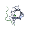
| Title | Human UBR-box from UBR2 in complex with HIFS peptide | ||||||
 Components Components |
| ||||||
 Keywords Keywords | LIGASE / UBR-box / N-end rule / Zinc finger / N-degron | ||||||
| Function / homology |  Function and homology information Function and homology informationhistone H2A ubiquitin ligase activity / L-leucine binding / ubiquitin-dependent protein catabolic process via the N-end rule pathway / male meiotic nuclear division / transposable element silencing / cellular response to L-leucine / positive regulation of T cell receptor signaling pathway / negative regulation of TOR signaling / reciprocal meiotic recombination / male meiosis I ...histone H2A ubiquitin ligase activity / L-leucine binding / ubiquitin-dependent protein catabolic process via the N-end rule pathway / male meiotic nuclear division / transposable element silencing / cellular response to L-leucine / positive regulation of T cell receptor signaling pathway / negative regulation of TOR signaling / reciprocal meiotic recombination / male meiosis I / protein K63-linked ubiquitination / ubiquitin ligase complex / RING-type E3 ubiquitin transferase / ubiquitin protein ligase activity / Antigen processing: Ubiquitination & Proteasome degradation / heterochromatin formation / spermatogenesis / proteasome-mediated ubiquitin-dependent protein catabolic process / protein ubiquitination / chromatin / zinc ion binding / nucleus / cytoplasm / cytosol Similarity search - Function | ||||||
| Biological species |  Homo sapiens (human) Homo sapiens (human) | ||||||
| Method |  X-RAY DIFFRACTION / SYNCHROTRON / MOLECULAR REPLACEMENT / Resolution: 1.55 Å X-RAY DIFFRACTION / SYNCHROTRON / MOLECULAR REPLACEMENT / Resolution: 1.55 Å | ||||||
 Authors Authors | Munoz-Escobar, J. / Kozlov, G. / Gehring, K. | ||||||
| Funding support |  Canada, 1items Canada, 1items
| ||||||
 Citation Citation | Journal: Structure / Year: 2017 Title: Bound Waters Mediate Binding of Diverse Substrates to a Ubiquitin Ligase. Authors: Munoz-Escobar, J. / Matta-Camacho, E. / Cho, C. / Kozlov, G. / Gehring, K. | ||||||
| History |
|
- Structure visualization
| Structure viewer | Molecule: MolmilJmol/JSmol |
|---|
- Downloads & links
-Download
| PDBx/mmCIF format | 5tdd.cif.gz | 59.5 KB | Display | PDBx/mmCIF format |
|---|---|---|---|---|
| PDB format | pdb5tdd.ent.gz | 42.3 KB | Display | PDB format |
| PDBx/mmJSON format | 5tdd.json.gz | Tree view | PDBx/mmJSON format | |
| Others |  Other downloads Other downloads |
-Validation report
| Arichive directory | https://data.pdbj.org/pub/pdb/validation_reports/td/5tddftp://data.pdbj.org/pub/pdb/validation_reports/td/5tdd | HTTPS FTP |
|---|
-Related structure data
| Related structure data |  5tdaC  5tdbC  5tdcC  5um3C  3ny3S C: citing same article ( S: Starting model for refinement |
|---|---|
| Similar structure data |


-Links
 PDBj
PDBj


- Assembly
| Deposited unit | 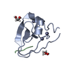
| ||||||||
|---|---|---|---|---|---|---|---|---|---|
| 1 |
| ||||||||
| Unit cell |
|
-Components
| #1: Protein | Mass: 8350.516 Da / Num. of mol.: 1 Source method: isolated from a genetically manipulated source Source: (gene. exp.) Homo sapiens (human) / Gene: UBR2, C6orf133, KIAA0349 / Plasmid: pGEX-6p-1 / Production host:  References: UniProt: Q8IWV8, Ligases; Forming carbon-nitrogen bonds; Acid-amino-acid ligases (peptide synthases) | ||||
|---|---|---|---|---|---|
| #2: Protein/peptide | Mass: 503.571 Da / Num. of mol.: 1 / Source method: obtained synthetically / Source: (synth.) Homo sapiens (human) | ||||
| #3: Chemical |   Mass: 65.409 Da / Num. of mol.: 3 / Source method: obtained synthetically / Formula: Zn Mass: 65.409 Da / Num. of mol.: 3 / Source method: obtained synthetically / Formula: Zn#4: Chemical |   Mass: 62.068 Da / Num. of mol.: 2 / Source method: obtained synthetically / Formula: C2H6O2 Mass: 62.068 Da / Num. of mol.: 2 / Source method: obtained synthetically / Formula: C2H6O2#5: Water | ChemComp-HOH / |  Mass: 18.015 Da / Num. of mol.: 44 / Source method: isolated from a natural source / Formula: H2O Mass: 18.015 Da / Num. of mol.: 44 / Source method: isolated from a natural source / Formula: H2O |
-Experimental details
-Experiment
| Experiment | Method: X-RAY DIFFRACTION / Number of used crystals: 1 |
|---|
- Sample preparation
| Crystal | Density Matthews: 1.8 Å3/Da / Density % sol: 30 % |
|---|---|
| Crystal grow | Temperature: 293 K / Method: vapor diffusion, sitting drop / pH: 6.5 / Details: 0.1 M MES pH 6.5, 12% PEG 20000 |
-Data collection
| Diffraction | Mean temperature: 100 K |
|---|---|
| Diffraction source | Source: SYNCHROTRON / Site: CHESS  / Beamline: A1 / Wavelength: 0.6362 Å / Beamline: A1 / Wavelength: 0.6362 Å |
| Detector | Type: ADSC QUANTUM 210 / Detector: CCD / Date: Mar 16, 2016 |
| Radiation | Protocol: SINGLE WAVELENGTH / Monochromatic (M) / Laue (L): M / Scattering type: x-ray |
| Radiation wavelength | Wavelength: 0.6362 Å / Relative weight: 1 |
| Reflection | Resolution: 1.55→20.727 Å / Num. obs: 9083 / % possible obs: 99.94 % / Redundancy: 6.4 % / Rsym value: 0.1 / Net I/σ(I): 32.1 |
| Reflection shell | Resolution: 1.55→1.58 Å / Redundancy: 5.7 % / Rmerge(I) obs: 0.558 / Mean I/σ(I) obs: 4.8 / CC1/2: 0.851 / % possible all: 100 |
- Processing
| Software |
| ||||||||||||||||||||||||||||||||||||||||||||||||||||||||
|---|---|---|---|---|---|---|---|---|---|---|---|---|---|---|---|---|---|---|---|---|---|---|---|---|---|---|---|---|---|---|---|---|---|---|---|---|---|---|---|---|---|---|---|---|---|---|---|---|---|---|---|---|---|---|---|---|---|
| Refinement | Method to determine structure: MOLECULAR REPLACEMENT Starting model: 3NY3 Resolution: 1.55→20.727 Å / SU ML: 0.11 / Cross valid method: THROUGHOUT / σ(F): 1.38 / Phase error: 14.01
| ||||||||||||||||||||||||||||||||||||||||||||||||||||||||
| Solvent computation | Shrinkage radii: 0.9 Å / VDW probe radii: 1.11 Å | ||||||||||||||||||||||||||||||||||||||||||||||||||||||||
| Refinement step | Cycle: LAST / Resolution: 1.55→20.727 Å
| ||||||||||||||||||||||||||||||||||||||||||||||||||||||||
| Refine LS restraints |
| ||||||||||||||||||||||||||||||||||||||||||||||||||||||||
| LS refinement shell |
|
