[English] 日本語
Yorodumi
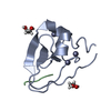
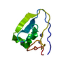
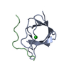
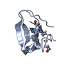
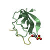
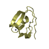
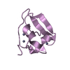
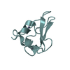
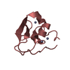
- PDB-3ny2: Structure of the ubr-box of UBR2 ubiquitin ligase -

+
Open data


ID or keywords:

Loading...

-
Basic information

Entry
Database: PDB / ID: 3ny2
TitleStructure of the ubr-box of UBR2 ubiquitin ligase
ComponentsE3 ubiquitin-protein ligase UBR2
KeywordsLIGASE / zinc finger-like / ubiquitin ligase
Function / homology
Function and homology information


leucine binding / ubiquitin-dependent protein catabolic process via the N-end rule pathway / histone ubiquitin ligase activity / male meiotic nuclear division / retrotransposon silencing / cellular response to leucine / negative regulation of TOR signaling / reciprocal meiotic recombination / male meiosis I / heterochromatin formation ...leucine binding / ubiquitin-dependent protein catabolic process via the N-end rule pathway / histone ubiquitin ligase activity / male meiotic nuclear division / retrotransposon silencing / cellular response to leucine / negative regulation of TOR signaling / reciprocal meiotic recombination / male meiosis I / heterochromatin formation / ubiquitin ligase complex / RING-type E3 ubiquitin transferase / ubiquitin protein ligase activity / Antigen processing: Ubiquitination & Proteasome degradation / spermatogenesis / protein ubiquitination / chromatin / zinc ion binding / nucleus / cytosol / cytoplasm
Similarity search - Function
: / E3 ubiquitin-protein ligase ELL-like / E3 ubiquitin-protein ligase UBR-like, C-terminal / Proteolysis_6 C-terminal / E3 ubiquitin-protein ligase UBR1-like / Putative zinc finger in N-recognin (UBR box) / Adaptor protein ClpS, core / ATP-dependent Clp protease adaptor protein ClpS / Zinc finger, UBR-type / Zinc finger UBR-type profile. ...: / E3 ubiquitin-protein ligase ELL-like / E3 ubiquitin-protein ligase UBR-like, C-terminal / Proteolysis_6 C-terminal / E3 ubiquitin-protein ligase UBR1-like / Putative zinc finger in N-recognin (UBR box) / Adaptor protein ClpS, core / ATP-dependent Clp protease adaptor protein ClpS / Zinc finger, UBR-type / Zinc finger UBR-type profile. / Putative zinc finger in N-recognin, a recognition component of the N-end rule pathway / Ribosomal protein L7/L12, C-terminal/adaptor protein ClpS-like / Winged helix DNA-binding domain superfamily
Similarity search - Domain/homology
E3 ubiquitin-protein ligase UBR2
Similarity search - Component
Biological speciesHomo sapiens (human)
MethodX-RAY DIFFRACTION / SYNCHROTRON / MOLECULAR REPLACEMENT / Resolution: 2.61 Å
AuthorsMatta-Camacho, E. / Kozlov, G. / Li, F. / Gehring, K.
CitationJournal: Nat.Struct.Mol.Biol. / Year: 2010
Title: Structural basis of substrate recognition and specificity in the N-end rule pathway.
Authors: Matta-Camacho, E. / Kozlov, G. / Li, F.F. / Gehring, K.
History
DepositionJul 14, 2010Deposition site: RCSB / Processing site: RCSB
Revision 1.0Aug 11, 2010Provider: repository / Type: Initial release
Revision 1.1Jul 13, 2011Group: Version format compliance
Revision 1.2Feb 21, 2024Group: Data collection / Database references ...Data collection / Database references / Derived calculations / Refinement description
Category: chem_comp_atom / chem_comp_bond ...chem_comp_atom / chem_comp_bond / database_2 / pdbx_struct_conn_angle / struct_conn / struct_ncs_dom_lim / struct_ref_seq_dif / struct_site
Item: _database_2.pdbx_DOI / _database_2.pdbx_database_accession ..._database_2.pdbx_DOI / _database_2.pdbx_database_accession / _pdbx_struct_conn_angle.ptnr1_auth_asym_id / _pdbx_struct_conn_angle.ptnr1_auth_comp_id / _pdbx_struct_conn_angle.ptnr1_auth_seq_id / _pdbx_struct_conn_angle.ptnr1_label_asym_id / _pdbx_struct_conn_angle.ptnr1_label_atom_id / _pdbx_struct_conn_angle.ptnr1_label_comp_id / _pdbx_struct_conn_angle.ptnr1_label_seq_id / _pdbx_struct_conn_angle.ptnr2_auth_asym_id / _pdbx_struct_conn_angle.ptnr2_auth_seq_id / _pdbx_struct_conn_angle.ptnr2_label_asym_id / _pdbx_struct_conn_angle.ptnr3_auth_asym_id / _pdbx_struct_conn_angle.ptnr3_auth_comp_id / _pdbx_struct_conn_angle.ptnr3_auth_seq_id / _pdbx_struct_conn_angle.ptnr3_label_asym_id / _pdbx_struct_conn_angle.ptnr3_label_atom_id / _pdbx_struct_conn_angle.ptnr3_label_comp_id / _pdbx_struct_conn_angle.ptnr3_label_seq_id / _pdbx_struct_conn_angle.value / _struct_conn.pdbx_dist_value / _struct_conn.ptnr1_auth_asym_id / _struct_conn.ptnr1_auth_comp_id / _struct_conn.ptnr1_auth_seq_id / _struct_conn.ptnr1_label_asym_id / _struct_conn.ptnr1_label_atom_id / _struct_conn.ptnr1_label_comp_id / _struct_conn.ptnr1_label_seq_id / _struct_conn.ptnr2_auth_asym_id / _struct_conn.ptnr2_auth_comp_id / _struct_conn.ptnr2_auth_seq_id / _struct_conn.ptnr2_label_asym_id / _struct_conn.ptnr2_label_atom_id / _struct_conn.ptnr2_label_comp_id / _struct_conn.ptnr2_label_seq_id / _struct_ncs_dom_lim.beg_auth_comp_id / _struct_ncs_dom_lim.beg_label_asym_id / _struct_ncs_dom_lim.beg_label_comp_id / _struct_ncs_dom_lim.beg_label_seq_id / _struct_ncs_dom_lim.end_auth_comp_id / _struct_ncs_dom_lim.end_label_asym_id / _struct_ncs_dom_lim.end_label_comp_id / _struct_ncs_dom_lim.end_label_seq_id / _struct_ref_seq_dif.details / _struct_site.pdbx_auth_asym_id / _struct_site.pdbx_auth_comp_id / _struct_site.pdbx_auth_seq_id

-
Structure visualization

Structure viewerMolecule:
MolmilJmol/JSmol

Downloads & links

-
Assembly

Deposited unit
A: E3 ubiquitin-protein ligase UBR2
B: E3 ubiquitin-protein ligase UBR2
C: E3 ubiquitin-protein ligase UBR2
D: E3 ubiquitin-protein ligase UBR2
E: E3 ubiquitin-protein ligase UBR2
F: E3 ubiquitin-protein ligase UBR2
G: E3 ubiquitin-protein ligase UBR2
H: E3 ubiquitin-protein ligase UBR2
hetero molecules


Theoretical massNumber of molelcules
Total (without water)67,46932
Polymers65,8998
Non-polymers1,57024
Water64936
1
A: E3 ubiquitin-protein ligase UBR2
hetero molecules


Theoretical massNumber of molelcules
Total (without water)8,4344
Polymers8,2371
Non-polymers1963
Water181
TypeNameSymmetry operationNumber
identity operation1_555x,y,z1
MethodPISA
2
B: E3 ubiquitin-protein ligase UBR2
hetero molecules


Theoretical massNumber of molelcules
Total (without water)8,4344
Polymers8,2371
Non-polymers1963
Water181
TypeNameSymmetry operationNumber
identity operation1_555x,y,z1
MethodPISA
3
C: E3 ubiquitin-protein ligase UBR2
hetero molecules


Theoretical massNumber of molelcules
Total (without water)8,4344
Polymers8,2371
Non-polymers1963
Water181
TypeNameSymmetry operationNumber
identity operation1_555x,y,z1
MethodPISA
4
D: E3 ubiquitin-protein ligase UBR2
hetero molecules


Theoretical massNumber of molelcules
Total (without water)8,4344
Polymers8,2371
Non-polymers1963
Water181
TypeNameSymmetry operationNumber
identity operation1_555x,y,z1
MethodPISA
5
E: E3 ubiquitin-protein ligase UBR2
hetero molecules


Theoretical massNumber of molelcules
Total (without water)8,4344
Polymers8,2371
Non-polymers1963
Water181
TypeNameSymmetry operationNumber
identity operation1_555x,y,z1
MethodPISA
6
F: E3 ubiquitin-protein ligase UBR2
hetero molecules


Theoretical massNumber of molelcules
Total (without water)8,4344
Polymers8,2371
Non-polymers1963
Water181
TypeNameSymmetry operationNumber
identity operation1_555x,y,z1
MethodPISA
7
G: E3 ubiquitin-protein ligase UBR2
hetero molecules


Theoretical massNumber of molelcules
Total (without water)8,4344
Polymers8,2371
Non-polymers1963
Water181
TypeNameSymmetry operationNumber
identity operation1_555x,y,z1
MethodPISA
8
H: E3 ubiquitin-protein ligase UBR2
hetero molecules


Theoretical massNumber of molelcules
Total (without water)8,4344
Polymers8,2371
Non-polymers1963
Water181
TypeNameSymmetry operationNumber
identity operation1_555x,y,z1
MethodPISA
Unit cell
Length a, b, c (Å)29.390, 61.456, 72.806
Angle α, β, γ (deg.)65.05, 89.98, 90.01
Int Tables number1
Space group name H-MP1
Noncrystallographic symmetry (NCS)NCS domain:
IDEns-IDDetails
11A
21B
31C
41D
51E
61F
71G
81H

NCS domain segments:

Component-ID: 1 / Ens-ID: 1 / Beg auth comp-ID: CYS / Beg label comp-ID: CYS / End auth comp-ID: HIS / End label comp-ID: HIS / Refine code: 3 / Auth seq-ID: 99 - 166 / Label seq-ID: 7 - 74

Dom-IDAuth asym-IDLabel asym-ID
1AA
2BB
3CC
4DD
5EE
6FF
7GG
8HH

-
Components

#1: Protein
E3 ubiquitin-protein ligase UBR2 / N-recognin-2 / Ubiquitin-protein ligase E3-alpha-2 / Ubiquitin-protein ligase E3-alpha-II


Mass: 8237.357 Da / Num. of mol.: 8 / Fragment: ubr-box
Source method: isolated from a genetically manipulated source
Source: (gene. exp.) Homo sapiens (human) / Gene: UBR2, C6orf133, KIAA0349 / Plasmid: pGEX-6P-1 / Production host: Escherichia coli (E. coli) / Strain (production host): BL21
References: UniProt: Q8IWV8, Ligases; Forming carbon-nitrogen bonds; Acid-amino-acid ligases (peptide synthases)
#2: Chemical...
ChemComp-ZN / ZINC ION


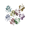
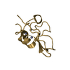
Mass: 65.409 Da / Num. of mol.: 24 / Source method: obtained synthetically / Formula: Zn
#3: Water ChemComp-HOH / water / Water


Mass: 18.015 Da / Num. of mol.: 36 / Source method: isolated from a natural source / Formula: H2O

-
Experimental details

-
Experiment

ExperimentMethod: X-RAY DIFFRACTION / Number of used crystals: 1

-
Sample preparation

CrystalDensity Matthews: 1.81 Å3/Da / Density % sol: 32.02 %
Crystal growTemperature: 293 K / Method: vapor diffusion, hanging drop / pH: 7
Details: 0.96M sodium citrate, pH 7.0, VAPOR DIFFUSION, HANGING DROP, temperature 293K

-
Data collection

DiffractionMean temperature: 100 K
Diffraction sourceSource: SYNCHROTRON / Site: CHESS / Beamline: A1 / Wavelength: 0.9779 Å
DetectorType: ADSC QUANTUM 210 / Detector: CCD / Date: Mar 4, 2010
RadiationMonochromator: Si 111 CHANNEL / Protocol: SINGLE WAVELENGTH / Monochromatic (M) / Laue (L): M / Scattering type: x-ray
Radiation wavelengthWavelength: 0.9779 Å / Relative weight: 1
ReflectionResolution: 2.6→50 Å / Num. all: 13189 / Num. obs: 12503 / % possible obs: 94.8 % / Observed criterion σ(F): 1 / Observed criterion σ(I): 1
Reflection shellResolution: 2.6→2.64 Å / % possible all: 79.1

-
Processing

Software
NameVersionClassification
ADSCQuantumdata collection
PHASERphasing
REFMAC5.2.0019refinement
HKL-2000data reduction
HKL-2000data scaling
RefinementMethod to determine structure: MOLECULAR REPLACEMENT / Resolution: 2.61→50 Å / Cor.coef. Fo:Fc: 0.927 / Cor.coef. Fo:Fc free: 0.886 / SU B: 15.629 / SU ML: 0.339 / Cross valid method: THROUGHOUT / σ(F): 1 / ESU R Free: 0.461 / Stereochemistry target values: MAXIMUM LIKELIHOOD
RfactorNum. reflection% reflectionSelection details
Rfree0.28808 652 5 %RANDOM
Rwork0.22974 ---
all0.24 13189 --
obs0.23265 12503 94.36 %-
Solvent computationIon probe radii: 0.8 Å / Shrinkage radii: 0.8 Å / VDW probe radii: 1.2 Å / Solvent model: MASK
Displacement parametersBiso mean: 32.62 Å2
Baniso -1Baniso -2Baniso -3
1--4.26 Å20.7 Å2-0.06 Å2
2--5.11 Å21.4 Å2
3----2.03 Å2
Refinement stepCycle: LAST / Resolution: 2.61→50 Å
ProteinNucleic acidLigandSolventTotal
Num. atoms4297 0 24 36 4357
Refine LS restraints
Refine-IDTypeDev idealDev ideal targetNumber
X-RAY DIFFRACTIONr_bond_refined_d0.0070.0214401
X-RAY DIFFRACTIONr_angle_refined_deg1.0941.9415934
X-RAY DIFFRACTIONr_dihedral_angle_1_deg5.4755567
X-RAY DIFFRACTIONr_dihedral_angle_2_deg33.28722.169189
X-RAY DIFFRACTIONr_dihedral_angle_3_deg17.75415664
X-RAY DIFFRACTIONr_dihedral_angle_4_deg21.7951534
X-RAY DIFFRACTIONr_chiral_restr0.0780.2601
X-RAY DIFFRACTIONr_gen_planes_refined0.0030.023450
X-RAY DIFFRACTIONr_nbd_refined0.1990.21774
X-RAY DIFFRACTIONr_nbtor_refined0.2950.22947
X-RAY DIFFRACTIONr_xyhbond_nbd_refined0.1410.2154
X-RAY DIFFRACTIONr_metal_ion_refined0.0830.211
X-RAY DIFFRACTIONr_symmetry_vdw_refined0.2380.2190
X-RAY DIFFRACTIONr_symmetry_hbond_refined0.20.218
X-RAY DIFFRACTIONr_mcbond_it0.3381.52895
X-RAY DIFFRACTIONr_mcangle_it0.60524466
X-RAY DIFFRACTIONr_scbond_it0.76831736
X-RAY DIFFRACTIONr_scangle_it1.2924.51468
Refine LS restraints NCS

Ens-ID: 1 / Refine-ID: X-RAY DIFFRACTION

Dom-IDAuth asym-IDNumberTypeRms dev position (Å)Weight position
1A240tight positional0.020.05
2B240tight positional0.020.05
3C240tight positional0.020.05
4D240tight positional0.020.05
5E240tight positional0.020.05
6F240tight positional0.020.05
7G240tight positional0.020.05
8H240tight positional0.020.05
1A205loose positional0.45
2B205loose positional0.375
3C205loose positional0.425
4D205loose positional0.555
5E205loose positional0.415
6F205loose positional0.395
7G205loose positional0.335
8H205loose positional0.415
1A240tight thermal0.040.5
2B240tight thermal0.040.5
3C240tight thermal0.030.5
4D240tight thermal0.040.5
5E240tight thermal0.040.5
6F240tight thermal0.030.5
7G240tight thermal0.030.5
8H240tight thermal0.030.5
1A205loose thermal0.6810
2B205loose thermal0.810
3C205loose thermal0.6910
4D205loose thermal0.6310
5E205loose thermal0.5810
6F205loose thermal0.6710
7G205loose thermal0.5910
8H205loose thermal0.6310
LS refinement shellResolution: 2.615→2.683 Å / Total num. of bins used: 20
RfactorNum. reflection% reflection
Rfree0.303 39 -
Rwork0.23 688 -
obs--72.19 %

+
About Yorodumi

-
News

-
Feb 9, 2022. New format data for meta-information of EMDB entries

New format data for meta-information of EMDB entries

  • Version 3 of the EMDB header file is now the official format.
  • The previous official version 1.9 will be removed from the archive.

Related info.:EMDB header

External links:wwPDB to switch to version 3 of the EMDB data model

-
Aug 12, 2020. Covid-19 info

Covid-19 info

URL: https://pdbj.org/emnavi/covid19.php

New page: Covid-19 featured information page in EM Navigator.

Related info.:Covid-19 info / Mar 5, 2020. Novel coronavirus structure data

+
Mar 5, 2020. Novel coronavirus structure data

Novel coronavirus structure data

Related info.:Yorodumi Speices / Aug 12, 2020. Covid-19 info

External links:COVID-19 featured content - PDBj / Molecule of the Month (242):Coronavirus Proteases

+
Jan 31, 2019. EMDB accession codes are about to change! (news from PDBe EMDB page)

EMDB accession codes are about to change! (news from PDBe EMDB page)

  • The allocation of 4 digits for EMDB accession codes will soon come to an end. Whilst these codes will remain in use, new EMDB accession codes will include an additional digit and will expand incrementally as the available range of codes is exhausted. The current 4-digit format prefixed with “EMD-” (i.e. EMD-XXXX) will advance to a 5-digit format (i.e. EMD-XXXXX), and so on. It is currently estimated that the 4-digit codes will be depleted around Spring 2019, at which point the 5-digit format will come into force.
  • The EM Navigator/Yorodumi systems omit the EMD- prefix.

Related info.:Q: What is EMD? / ID/Accession-code notation in Yorodumi/EM Navigator

External links:EMDB Accession Codes are Changing Soon! / Contact to PDBj

+
Jul 12, 2017. Major update of PDB

Major update of PDB

  • wwPDB released updated PDB data conforming to the new PDBx/mmCIF dictionary.
  • This is a major update changing the version number from 4 to 5, and with Remediation, in which all the entries are updated.
  • In this update, many items about electron microscopy experimental information are reorganized (e.g. em_software).
  • Now, EM Navigator and Yorodumi are based on the updated data.

External links:wwPDB Remediation / Enriched Model Files Conforming to OneDep Data Standards Now Available in the PDB FTP Archive

-
Yorodumi

Thousand views of thousand structures

  • Yorodumi is a browser for structure data from EMDB, PDB, SASBDB, etc.
  • This page is also the successor to EM Navigator detail page, and also detail information page/front-end page for Omokage search.
  • The word "yorodu" (or yorozu) is an old Japanese word meaning "ten thousand". "mi" (miru) is to see.

Related info.:EMDB / PDB / SASBDB / Comparison of 3 databanks / Yorodumi Search / Aug 31, 2016. New EM Navigator & Yorodumi / Yorodumi Papers / Jmol/JSmol / Function and homology information / Changes in new EM Navigator and Yorodumi

Read more