 Movie
Movie Controller

[English] 日本語
 Yorodumi
- PDB-5t01: Human c-Jun DNA binding domain homodimer in complex with methylat... -
+ Open data

- Basic information
| Entry | Database: PDB / ID: 5t01 | ||||||
|---|---|---|---|---|---|---|---|
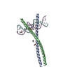
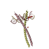
| Title | Human c-Jun DNA binding domain homodimer in complex with methylated DNA | ||||||
 Components Components |
| ||||||
 Keywords Keywords | TRANSCRIPTION REGULATOR/DNA / Zta / Zebra / BZLF-1 / AP-1 / Epstein-Barr virus / EBV / 5-methylcytosine / 5mC / DNA methylation / transcription factor / basic leucine-zipper / bZIP / TRANSCRIPTION REGULATOR-DNA complex | ||||||
| Function / homology |  Function and homology information Function and homology informationleading edge cell differentiation / cellular response to anisomycin / cAMP response element binding / transcription factor AP-1 complex / integrated stress response signaling / negative regulation of DNA binding / release from viral latency / WNT5:FZD7-mediated leishmania damping / SMAD protein signal transduction / host-mediated activation of viral transcription ...leading edge cell differentiation / cellular response to anisomycin / cAMP response element binding / transcription factor AP-1 complex / integrated stress response signaling / negative regulation of DNA binding / release from viral latency / WNT5:FZD7-mediated leishmania damping / SMAD protein signal transduction / host-mediated activation of viral transcription / response to steroid hormone / axon regeneration / positive regulation of DNA-templated transcription initiation / Deregulated CDK5 triggers multiple neurodegenerative pathways in Alzheimer's disease models / Activation of the AP-1 family of transcription factors / nuclear chromosome / eyelid development in camera-type eye / outflow tract morphogenesis / R-SMAD binding / positive regulation of epithelial cell migration / ubiquitin-like protein ligase binding / host-mediated suppression of viral transcription / monocyte differentiation / general transcription initiation factor binding / positive regulation of vascular associated smooth muscle cell proliferation / JNK cascade / response to muscle stretch / transcription repressor complex / positive regulation of endothelial cell proliferation / transforming growth factor beta receptor signaling pathway / cellular response to calcium ion / response to endoplasmic reticulum stress / Regulation of PTEN gene transcription / GTPase activator activity / FCERI mediated MAPK activation / TP53 Regulates Transcription of DNA Repair Genes / liver development / euchromatin / microglial cell activation / positive regulation of miRNA transcription / MAPK6/MAPK4 signaling / DNA-binding transcription repressor activity, RNA polymerase II-specific / Pre-NOTCH Transcription and Translation / Activation of anterior HOX genes in hindbrain development during early embryogenesis / RNA polymerase II transcription regulator complex / positive regulation of fibroblast proliferation / sequence-specific double-stranded DNA binding / Signaling by ALK fusions and activated point mutants / regulation of cell population proliferation / Senescence-Associated Secretory Phenotype (SASP) / angiogenesis / DNA-binding transcription activator activity, RNA polymerase II-specific / transcription regulator complex / Oxidative Stress Induced Senescence / Estrogen-dependent gene expression / RNA polymerase II-specific DNA-binding transcription factor binding / negative regulation of neuron apoptotic process / DNA-binding transcription factor activity, RNA polymerase II-specific / positive regulation of ERK1 and ERK2 cascade / cell population proliferation / regulation of cell cycle / transcription cis-regulatory region binding / RNA polymerase II cis-regulatory region sequence-specific DNA binding / DNA-binding transcription factor activity / response to xenobiotic stimulus / negative regulation of cell population proliferation / negative regulation of DNA-templated transcription / apoptotic process / chromatin binding / ubiquitin protein ligase binding / regulation of transcription by RNA polymerase II / positive regulation of DNA-templated transcription / chromatin / enzyme binding / negative regulation of transcription by RNA polymerase II / positive regulation of transcription by RNA polymerase II / DNA binding / RNA binding / nucleoplasm / identical protein binding / nucleus Similarity search - Function | ||||||
| Biological species |  Homo sapiens (human) Homo sapiens (human)synthetic construct (others) | ||||||
| Method |  X-RAY DIFFRACTION / SYNCHROTRON / MOLECULAR REPLACEMENT / Resolution: 1.89 Å X-RAY DIFFRACTION / SYNCHROTRON / MOLECULAR REPLACEMENT / Resolution: 1.89 Å | ||||||
 Authors Authors | Hong, S. / Horton, J.R. / Cheng, X. | ||||||
| Funding support |  United States, 1items United States, 1items
| ||||||
 Citation Citation | Journal: Nucleic Acids Res. / Year: 2017 Title: Methyl-dependent and spatial-specific DNA recognition by the orthologous transcription factors human AP-1 and Epstein-Barr virus Zta. Authors: Hong, S. / Wang, D. / Horton, J.R. / Zhang, X. / Speck, S.H. / Blumenthal, R.M. / Cheng, X. | ||||||
| History |
|
- Structure visualization
| Structure viewer | Molecule: MolmilJmol/JSmol |
|---|
- Downloads & links
-Download
| PDBx/mmCIF format | 5t01.cif.gz | 110.2 KB | Display | PDBx/mmCIF format |
|---|---|---|---|---|
| PDB format | pdb5t01.ent.gz | 82.1 KB | Display | PDB format |
| PDBx/mmJSON format | 5t01.json.gz | Tree view | PDBx/mmJSON format | |
| Others |  Other downloads Other downloads |
-Validation report
| Arichive directory | https://data.pdbj.org/pub/pdb/validation_reports/t0/5t01ftp://data.pdbj.org/pub/pdb/validation_reports/t0/5t01 | HTTPS FTP |
|---|
-Related structure data
| Related structure data | 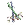 5szxC 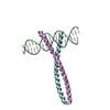 1fosS C: citing same article ( S: Starting model for refinement |
|---|---|
| Similar structure data |


-Links
 PDBj
PDBj


- Assembly
| Deposited unit | 
| ||||||||
|---|---|---|---|---|---|---|---|---|---|
| 1 |
| ||||||||
| Unit cell |
|
-Components
| #1: DNA chain | Mass: 5706.697 Da / Num. of mol.: 1 / Source method: obtained synthetically / Source: (synth.) synthetic construct (others) | ||
|---|---|---|---|
| #2: DNA chain | Mass: 5956.901 Da / Num. of mol.: 1 / Source method: obtained synthetically / Source: (synth.) synthetic construct (others) | ||
| #3: Protein | Mass: 7591.038 Da / Num. of mol.: 2 / Fragment: DNA binding domain (UNP residues 254-315) / Mutation: C269S Source method: isolated from a genetically manipulated source Source: (gene. exp.) Homo sapiens (human) / Gene: JUN / Production host:  #4: Water | ChemComp-HOH / |  Mass: 18.015 Da / Num. of mol.: 120 / Source method: isolated from a natural source / Formula: H2O Mass: 18.015 Da / Num. of mol.: 120 / Source method: isolated from a natural source / Formula: H2O |
-Experimental details
-Experiment
| Experiment | Method: X-RAY DIFFRACTION / Number of used crystals: 1 |
|---|
- Sample preparation
| Crystal | Density Matthews: 2.81 Å3/Da / Density % sol: 56.25 % |
|---|---|
| Crystal grow | Temperature: 289 K / Method: vapor diffusion, hanging drop / pH: 5 Details: 0.05 M Citric acid 0.05 M Bis-Tris propane 16% PEG3350 |
-Data collection
| Diffraction | Mean temperature: 100 K |
|---|---|
| Diffraction source | Source: SYNCHROTRON / Site: APS / Beamline: 22-ID / Wavelength: 1 Å |
| Detector | Type: RAYONIX MX300-HS / Detector: CCD / Date: Feb 27, 2015 |
| Radiation | Protocol: SINGLE WAVELENGTH / Monochromatic (M) / Laue (L): M / Scattering type: x-ray |
| Radiation wavelength | Wavelength: 1 Å / Relative weight: 1 |
| Reflection | Resolution: 1.89→35 Å / Num. obs: 23735 / % possible obs: 98.5 % / Redundancy: 6.9 % / Rmerge(I) obs: 0.083 / Net I/σ(I): 20.3 |
| Reflection shell | Resolution: 1.89→1.96 Å / Redundancy: 4.2 % / Rmerge(I) obs: 0.8 / Mean I/σ(I) obs: 2.63 / % possible all: 90.1 |
- Processing
| Software |
| |||||||||||||||||||||||||||||||||||||||||||||||||||||||||||||||
|---|---|---|---|---|---|---|---|---|---|---|---|---|---|---|---|---|---|---|---|---|---|---|---|---|---|---|---|---|---|---|---|---|---|---|---|---|---|---|---|---|---|---|---|---|---|---|---|---|---|---|---|---|---|---|---|---|---|---|---|---|---|---|---|---|
| Refinement | Method to determine structure: MOLECULAR REPLACEMENT Starting model: 1FOS Resolution: 1.89→27.75 Å / SU ML: 0.24 / Cross valid method: FREE R-VALUE / σ(F): 1.35 / Phase error: 28.09 / Stereochemistry target values: ML
| |||||||||||||||||||||||||||||||||||||||||||||||||||||||||||||||
| Solvent computation | Shrinkage radii: 0.9 Å / VDW probe radii: 1.11 Å / Solvent model: FLAT BULK SOLVENT MODEL | |||||||||||||||||||||||||||||||||||||||||||||||||||||||||||||||
| Refinement step | Cycle: LAST / Resolution: 1.89→27.75 Å
| |||||||||||||||||||||||||||||||||||||||||||||||||||||||||||||||
| Refine LS restraints |
| |||||||||||||||||||||||||||||||||||||||||||||||||||||||||||||||
| LS refinement shell |
| |||||||||||||||||||||||||||||||||||||||||||||||||||||||||||||||
| Refinement TLS params. | Method: refined / Origin x: -24.4193 Å / Origin y: 16.7252 Å / Origin z: 20.1421 Å
| |||||||||||||||||||||||||||||||||||||||||||||||||||||||||||||||
| Refinement TLS group | Selection details: all |
