 Movie
Movie Controller

[English] 日本語
 Yorodumi
- PDB-5szk: Structure of human N-terminally engineered Rab1b in complex with ... -
+ Open data

- Basic information
| Entry | Database: PDB / ID: 5szk | |||||||||
|---|---|---|---|---|---|---|---|---|---|---|
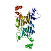
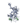
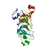
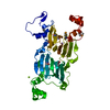
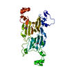
| Title | Structure of human N-terminally engineered Rab1b in complex with the bMERB domain of Mical-cL | |||||||||
 Components Components |
| |||||||||
 Keywords Keywords | ENDOCYTOSIS / Mical-cL / DUF3585 / Mical / Rab effector / Rab1b / protein transport | |||||||||
| Function / homology |  Function and homology information Function and homology informationpositive regulation of glycoprotein metabolic process / F-actin monooxygenase / F-actin monooxygenase activity / sulfur oxidation / NAD(P)H oxidase H2O2-forming activity / oxidoreductase activity, acting on paired donors, with incorporation or reduction of molecular oxygen, NAD(P)H as one donor, and incorporation of one atom of oxygen / phagophore assembly site membrane / RAB geranylgeranylation / regulation of autophagosome assembly / RAB GEFs exchange GTP for GDP on RABs ...positive regulation of glycoprotein metabolic process / F-actin monooxygenase / F-actin monooxygenase activity / sulfur oxidation / NAD(P)H oxidase H2O2-forming activity / oxidoreductase activity, acting on paired donors, with incorporation or reduction of molecular oxygen, NAD(P)H as one donor, and incorporation of one atom of oxygen / phagophore assembly site membrane / RAB geranylgeranylation / regulation of autophagosome assembly / RAB GEFs exchange GTP for GDP on RABs / actin filament depolymerization / Golgi Cisternae Pericentriolar Stack Reorganization / mitogen-activated protein kinase binding / COPII-mediated vesicle transport / COPI-dependent Golgi-to-ER retrograde traffic / virion assembly / heart looping / Golgi organization / endoplasmic reticulum to Golgi vesicle-mediated transport / autophagosome assembly / transport vesicle / COPI-mediated anterograde transport / FAD binding / endoplasmic reticulum-Golgi intermediate compartment membrane / cytoskeleton organization / endomembrane system / small monomeric GTPase / actin filament / monooxygenase activity / intracellular protein transport / heart development / actin binding / G protein activity / cytoskeleton / oxidoreductase activity / Golgi membrane / GTPase activity / endoplasmic reticulum membrane / GTP binding / perinuclear region of cytoplasm / Golgi apparatus / positive regulation of transcription by RNA polymerase II / extracellular exosome / metal ion binding / nucleus / cytoplasm / cytosol Similarity search - Function | |||||||||
| Biological species |  Homo sapiens (human) Homo sapiens (human) | |||||||||
| Method |  X-RAY DIFFRACTION / SYNCHROTRON / MOLECULAR REPLACEMENT / Resolution: 2.8 Å X-RAY DIFFRACTION / SYNCHROTRON / MOLECULAR REPLACEMENT / Resolution: 2.8 Å | |||||||||
 Authors Authors | Rai, A. / Oprisko, A. / Campos, J. / Fu, Y. / Friese, T. / Itzen, A. / Goody, R.S. / Gazdag, E.M. / Mueller, M.P. | |||||||||
| Funding support |  Germany, 2items Germany, 2items
| |||||||||
 Citation Citation | Journal: Elife / Year: 2016 Title: bMERB domains are bivalent Rab8 family effectors evolved by gene duplication. Authors: Rai, A. / Oprisko, A. / Campos, J. / Fu, Y. / Friese, T. / Itzen, A. / Goody, R.S. / Gazdag, E.M. / Muller, M.P. | |||||||||
| History |
|
- Structure visualization
| Structure viewer | Molecule: MolmilJmol/JSmol |
|---|
- Downloads & links
-Download
| PDBx/mmCIF format | 5szk.cif.gz | 147.6 KB | Display | PDBx/mmCIF format |
|---|---|---|---|---|
| PDB format | pdb5szk.ent.gz | 116 KB | Display | PDB format |
| PDBx/mmJSON format | 5szk.json.gz | Tree view | PDBx/mmJSON format | |
| Others |  Other downloads Other downloads |
-Validation report
| Arichive directory | https://data.pdbj.org/pub/pdb/validation_reports/sz/5szkftp://data.pdbj.org/pub/pdb/validation_reports/sz/5szk | HTTPS FTP |
|---|
-Related structure data
| Related structure data | 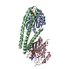 5lpnC 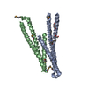 5szgSC 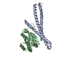 5szhC 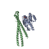 5sziC  5szjC 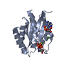 3nkvS S: Starting model for refinement C: citing same article ( |
|---|---|
| Similar structure data |


-Links
 PDBj
PDBj


- Assembly
| Deposited unit | 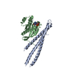
| ||||||||
|---|---|---|---|---|---|---|---|---|---|
| 1 |
| ||||||||
| Unit cell |
|
-Components
| #1: Protein | Mass: 18469.699 Da / Num. of mol.: 1 Source method: isolated from a genetically manipulated source Source: (gene. exp.) Homo sapiens (human) / Gene: MICALCL / Production host:  |
|---|---|
| #2: Protein | Mass: 22352.361 Da / Num. of mol.: 1 / Mutation: N2A, P3K, E4T Source method: isolated from a genetically manipulated source Source: (gene. exp.) Homo sapiens (human) / Gene: RAB1B / Production host: |
| #3: Chemical | ChemComp-MG /   Mass: 24.305 Da / Num. of mol.: 1 / Mutation: N2A, P3K, E4T Mass: 24.305 Da / Num. of mol.: 1 / Mutation: N2A, P3K, E4TSource method: isolated from a genetically manipulated source Formula: Mg / Source: (gene. exp.) Homo sapiens (human) / Production host: |
| #4: Chemical | ChemComp-GNP /   Mass: 522.196 Da / Num. of mol.: 1 / Mutation: N2A, P3K, E4T Mass: 522.196 Da / Num. of mol.: 1 / Mutation: N2A, P3K, E4TSource method: isolated from a genetically manipulated source Formula: C10H17N6O13P3 / Source: (gene. exp.) Homo sapiens (human) / Production host: Comment: GppNHp, GMPPNP, energy-carrying molecule analogue*YM |
| #5: Water | ChemComp-HOH /  Mass: 18.015 Da / Num. of mol.: 3 / Source method: isolated from a natural source / Formula: H2O Mass: 18.015 Da / Num. of mol.: 3 / Source method: isolated from a natural source / Formula: H2O |
-Experimental details
-Experiment
| Experiment | Method: X-RAY DIFFRACTION / Number of used crystals: 1 |
|---|
- Sample preparation
| Crystal | Density Matthews: 3.07 Å3/Da / Density % sol: 59.95 % |
|---|---|
| Crystal grow | Temperature: 293 K / Method: vapor diffusion, hanging drop / pH: 7.5 Details: 0.1M bis-tris pH 7.5, 0.2M sodium malonate and 20% (w/v) PEG3350 |
-Data collection
| Diffraction | Mean temperature: 100 K |
|---|---|
| Diffraction source | Source: SYNCHROTRON / Site: SLS  / Beamline: X10SA / Wavelength: 0.91908 Å / Beamline: X10SA / Wavelength: 0.91908 Å |
| Detector | Type: DECTRIS PILATUS 6M / Detector: PIXEL / Date: Oct 26, 2015 |
| Radiation | Monochromator: SI(111) / Protocol: SINGLE WAVELENGTH / Monochromatic (M) / Laue (L): M / Scattering type: x-ray |
| Radiation wavelength | Wavelength: 0.91908 Å / Relative weight: 1 |
| Reflection | Resolution: 2.8→44.8 Å / Num. obs: 12904 / % possible obs: 100 % / Redundancy: 19.2 % / Rmerge(I) obs: 0.076 / Rsym value: 0.079 / Net I/σ(I): 22.8 |
| Reflection shell | Resolution: 2.8→2.85 Å / Redundancy: 13.8 % / Rmerge(I) obs: 1.11 / Mean I/σ(I) obs: 2.45 / CC1/2: 0.757 / % possible all: 100 |
- Processing
| Software |
| |||||||||||||||||||||||||||||||||||||||||||||||||||||||||||||||||||||||||||
|---|---|---|---|---|---|---|---|---|---|---|---|---|---|---|---|---|---|---|---|---|---|---|---|---|---|---|---|---|---|---|---|---|---|---|---|---|---|---|---|---|---|---|---|---|---|---|---|---|---|---|---|---|---|---|---|---|---|---|---|---|---|---|---|---|---|---|---|---|---|---|---|---|---|---|---|---|
| Refinement | Method to determine structure: MOLECULAR REPLACEMENT Starting model: 3NKV, 5SZG Resolution: 2.8→44.8 Å / SU ML: 0.46 / Cross valid method: FREE R-VALUE / σ(F): 1.36 / Phase error: 30.44
| |||||||||||||||||||||||||||||||||||||||||||||||||||||||||||||||||||||||||||
| Solvent computation | Shrinkage radii: 0.9 Å / VDW probe radii: 1.11 Å | |||||||||||||||||||||||||||||||||||||||||||||||||||||||||||||||||||||||||||
| Refinement step | Cycle: LAST / Resolution: 2.8→44.8 Å
| |||||||||||||||||||||||||||||||||||||||||||||||||||||||||||||||||||||||||||
| Refine LS restraints |
| |||||||||||||||||||||||||||||||||||||||||||||||||||||||||||||||||||||||||||
| LS refinement shell |
| |||||||||||||||||||||||||||||||||||||||||||||||||||||||||||||||||||||||||||
| Refinement TLS params. | Method: refined / Refine-ID: X-RAY DIFFRACTION
| |||||||||||||||||||||||||||||||||||||||||||||||||||||||||||||||||||||||||||
| Refinement TLS group |
|
