F-actin monooxygenase / F-actin monooxygenase activity / oxidoreductase activity, acting on paired donors, with incorporation or reduction of molecular oxygen, NAD(P)H as one donor, and incorporation of one atom of oxygen / actin filament depolymerization / Flemming body / exocytosis / cytoskeleton organization / FAD binding / spindle / actin cytoskeleton organization ...F-actin monooxygenase / F-actin monooxygenase activity / oxidoreductase activity, acting on paired donors, with incorporation or reduction of molecular oxygen, NAD(P)H as one donor, and incorporation of one atom of oxygen / actin filament depolymerization / Flemming body / exocytosis / cytoskeleton organization / FAD binding / spindle / actin cytoskeleton organization / actin binding / cell cortex / molecular adaptor activity / cell division / nucleoplasm / metal ion binding / nucleus / plasma membrane / cytosol Similarity search - Function
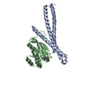
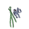
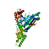
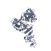
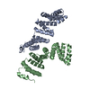
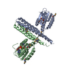
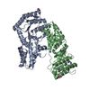
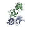
Protein-methioninesulfoxideoxidaseMICAL3 / Molecule interacting with CasL protein 3 / MICAL-3
Mass: 18537.062 Da / Num. of mol.: 2 / Fragment: UNP residues 1841-1990 Source method: isolated from a genetically manipulated source Source: (gene. exp.) Homo sapiens (human) / Gene: MICAL3, KIAA0819, KIAA1364 / Production host: Escherichia coli (E. coli) References: UniProt: Q7RTP6, Oxidoreductases; Acting on paired donors, with incorporation or reduction of molecular oxygen; With NADH or NADPH as one donor, and incorporation of one atom of oxygen into the other donor
Type: MARMOSAIC 225 mm CCD / Detector: CCD / Date: Dec 11, 2009
Radiation
Monochromator: SI(111) / Protocol: SINGLE WAVELENGTH / Monochromatic (M) / Laue (L): M / Scattering type: x-ray
Radiation wavelength
Wavelength: 0.978956 Å / Relative weight: 1
Reflection
Resolution: 2.7→47.8 Å / Num. obs: 20544 / % possible obs: 99.1 % / Redundancy: 13.3 % / Rmerge(I) obs: 0.138 / Rsym value: 0.144 / Net I/σ(I): 16.8
Reflection shell
Resolution: 2.7→2.8 Å / Redundancy: 13.6 % / Rmerge(I) obs: 1.15 / Mean I/σ(I) obs: 3.6 / CC1/2: 0.87 / % possible all: 98.5
-
Processing
Software
Name
Version
Classification
REFMAC
5.8.0103
refinement
XDS
datareduction
XSCALE
datascaling
PHASER
phasing
Refinement
Method to determine structure: SAD / Resolution: 2.7→47.78 Å / Cor.coef. Fo:Fc: 0.923 / Cor.coef. Fo:Fc free: 0.901 / SU B: 8.62 / SU ML: 0.19 / Cross valid method: THROUGHOUT / ESU R: 0.691 / ESU R Free: 0.348 / Details: HYDROGENS HAVE BEEN ADDED IN THE RIDING POSITIONS
Rfactor
Num. reflection
% reflection
Selection details
Rfree
0.28208
556
5 %
RANDOM
Rwork
0.2509
-
-
-
obs
0.25245
10553
98.83 %
-
Solvent computation
Ion probe radii: 0.8 Å / Shrinkage radii: 0.8 Å / VDW probe radii: 1.2 Å
 Movie
Movie Controller


 Open data
 Basic information
 Components
 Keywords
 Function and homology information
 Homo sapiens (human)
 X-RAY DIFFRACTION /
  Authors
 Germany, 2items
  Citation
 Structure visualization
 Downloads & links
 Other downloads
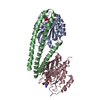


 PDBj
PDBj Assembly


 Mass: 106.120 Da / Num. of mol.: 2 / Source method: obtained synthetically / Formula: C4H10O3
 Sample preparation
 / Beamline: X10SA / Wavelength: 0.978956 Å
 Processing