 Movie
Movie Controller


+ Open data

- Basic information
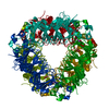
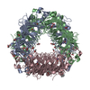
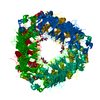
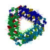
| Entry | Database: PDB / ID: 1uw8 | ||||||
|---|---|---|---|---|---|---|---|
| Title | CRYSTAL STRUCTURE OF OXALATE DECARBOXYLASE | ||||||
 Components Components | OXALATE DECARBOXYLASE OXDC | ||||||
 Keywords Keywords | LYASE / METAL BINDING PROTEIN / CUPIN / DECARBOXYLASE / OXALATE / MANGANESE / FORMATE | ||||||
| Function / homology |  Function and homology information Function and homology informationoxalate decarboxylase / oxalate decarboxylase activity / oxalate metabolic process / metal ion binding / cytoplasm Similarity search - Function | ||||||
| Biological species |  | ||||||
| Method |  X-RAY DIFFRACTION / MOLECULAR REPLACEMENT / Resolution: 2 Å X-RAY DIFFRACTION / MOLECULAR REPLACEMENT / Resolution: 2 Å | ||||||
 Authors Authors | Just, V.J. / Stevenson, C.E.M. / Bowater, L. / Tanner, A. / Lawson, D.M. / Bornemann, S. | ||||||
 Citation Citation | Journal: J.Biol.Chem. / Year: 2004 Title: A Closed Conformation of Bacillus Subtilis Oxalate Decarboxylase Oxdc Provides Evidence for the True Identity of the Active Site Authors: Just, V.J. / Stevenson, C.E.M. / Bowater, L. / Tanner, A. / Lawson, D.M. / Bornemann, S. #1: Journal: Biochemistry / Year: 2002Title: Structure of Oxalate Decarboxylase from Bacillus Subtilis at 1.75 A Resolution Authors: Anand, R. / Dorrestein, P.C. / Kinsland, C. / Begley, T.P. / Ealick, S.E. #2: Journal: J.Biol.Chem. / Year: 2001 Title: Oxalate Decarboxylase Requires Manganese and Dioxygen for Activity Authors: Tanner, A. / Bowater, L. / Fairhurst, S.A. / Bornemann, S. | ||||||
| History |
| ||||||
| Remark 700 | SHEET THE SHEET STRUCTURE OF THIS MOLECULE IS BIFURCATED. IN ORDER TO REPRESENT THIS FEATURE IN ... SHEET THE SHEET STRUCTURE OF THIS MOLECULE IS BIFURCATED. IN ORDER TO REPRESENT THIS FEATURE IN THE SHEET RECORDS BELOW, TWO SHEETS ARE DEFINED. |
- Structure visualization
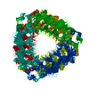
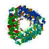
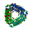
| Structure viewer | Molecule: MolmilJmol/JSmol |
|---|
- Downloads & links
-Download
| PDBx/mmCIF format | 1uw8.cif.gz | 97.7 KB | Display | PDBx/mmCIF format |
|---|---|---|---|---|
| PDB format | pdb1uw8.ent.gz | 73 KB | Display | PDB format |
| PDBx/mmJSON format | 1uw8.json.gz | Tree view | PDBx/mmJSON format | |
| Others |  Other downloads Other downloads |
-Validation report
| Arichive directory | https://data.pdbj.org/pub/pdb/validation_reports/uw/1uw8ftp://data.pdbj.org/pub/pdb/validation_reports/uw/1uw8 | HTTPS FTP |
|---|
-Related structure data
| Related structure data | 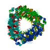 1j58S S: Starting model for refinement |
|---|---|
| Similar structure data |


-Links
 PDBj
PDBj- Assembly
| Deposited unit | 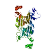
| ||||||||
|---|---|---|---|---|---|---|---|---|---|
| 1 | x 6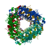
| ||||||||
| Unit cell |
| ||||||||
| Components on special symmetry positions |
|
-Components
| #1: Protein | Mass: 43620.902 Da / Num. of mol.: 1 Source method: isolated from a genetically manipulated source Details: FORMERLY KNOWN AS YVRK / Source: (gene. exp.) | ||||
|---|---|---|---|---|---|
| #2: Chemical |   Mass: 54.938 Da / Num. of mol.: 2 / Source method: obtained synthetically / Formula: Mn Mass: 54.938 Da / Num. of mol.: 2 / Source method: obtained synthetically / Formula: Mn#3: Chemical | ChemComp-TRS / |   Mass: 122.143 Da / Num. of mol.: 1 / Source method: obtained synthetically / Formula: C4H12NO3 / Comment: pH buffer*YM Mass: 122.143 Da / Num. of mol.: 1 / Source method: obtained synthetically / Formula: C4H12NO3 / Comment: pH buffer*YM#4: Water | ChemComp-HOH / |  Mass: 18.015 Da / Num. of mol.: 402 / Source method: isolated from a natural source / Formula: H2O Mass: 18.015 Da / Num. of mol.: 402 / Source method: isolated from a natural source / Formula: H2O |
-Experimental details
-Experiment
| Experiment | Method: X-RAY DIFFRACTION / Number of used crystals: 1 |
|---|
- Sample preparation
| Crystal | Density Matthews: 3.2 Å3/Da / Density % sol: 62 % | ||||||||||||||||||||||||||||||
|---|---|---|---|---|---|---|---|---|---|---|---|---|---|---|---|---|---|---|---|---|---|---|---|---|---|---|---|---|---|---|---|
| Crystal grow | Temperature: 291 K / pH: 8.5 / Details: 8% PEG 8000, TRIS-HCL PH8.5, 18 DEG C, pH 8.50 | ||||||||||||||||||||||||||||||
| Crystal grow | *PLUS Temperature: 18 ℃ / pH: 7 / Method: vapor diffusion, hanging drop | ||||||||||||||||||||||||||||||
| Components of the solutions | *PLUS
|
-Data collection
| Diffraction | Mean temperature: 100 K |
|---|---|
| Diffraction source | Source: ROTATING ANODE / Type: RIGAKU RUH3R / Wavelength: 1.5418 |
| Detector | Type: MARRESEARCH / Detector: IMAGE PLATE / Date: Sep 21, 2001 / Details: MIRRORS |
| Radiation | Protocol: SINGLE WAVELENGTH / Monochromatic (M) / Laue (L): M / Scattering type: x-ray |
| Radiation wavelength | Wavelength: 1.5418 Å / Relative weight: 1 |
| Reflection | Resolution: 2→40 Å / Num. obs: 38069 / % possible obs: 100 % / Observed criterion σ(I): -3 / Redundancy: 23.6 % / Rmerge(I) obs: 0.084 / Net I/σ(I): 34.4 |
| Reflection shell | Resolution: 2→2.03 Å / Rmerge(I) obs: 0.328 / Mean I/σ(I) obs: 6.7 / % possible all: 99.8 |
| Reflection | *PLUS Highest resolution: 2 Å / Lowest resolution: 40 Å / Redundancy: 23.6 % / Rmerge(I) obs: 0.084 |
| Reflection shell | *PLUS % possible obs: 99.8 % / Rmerge(I) obs: 0.328 / Mean I/σ(I) obs: 6.7 |
- Processing
| Software |
| ||||||||||||||||||||
|---|---|---|---|---|---|---|---|---|---|---|---|---|---|---|---|---|---|---|---|---|---|
| Refinement | Method to determine structure: MOLECULAR REPLACEMENT Starting model: PDB ENTRY 1J58 Resolution: 2→91.29 Å / SU B: 1.95 / SU ML: 0.056 / Cross valid method: THROUGHOUT / ESU R: 0.101 / ESU R Free: 0.099
| ||||||||||||||||||||
| Displacement parameters | Biso mean: 16.62 Å2
| ||||||||||||||||||||
| Refinement step | Cycle: LAST / Resolution: 2→91.29 Å
| ||||||||||||||||||||
| Refinement | *PLUS % reflection Rfree: 5 % / Rfactor Rfree: 0.158 / Rfactor Rwork: 0.127 | ||||||||||||||||||||
| Solvent computation | *PLUS | ||||||||||||||||||||
| Displacement parameters | *PLUS | ||||||||||||||||||||
| Refine LS restraints | *PLUS
|
