 Movie
Movie Controller

[English] 日本語
 Yorodumi
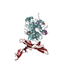
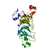
- PDB-3zld: Crystal structure of Toxoplasma gondii sporozoite AMA1 in complex... -
+ Open data

- Basic information
| Entry | Database: PDB / ID: 3zld | ||||||
|---|---|---|---|---|---|---|---|
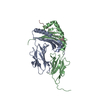
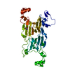
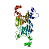
| Title | Crystal structure of Toxoplasma gondii sporozoite AMA1 in complex with a 36 aa region of sporozoite RON2 | ||||||
 Components Components |
| ||||||
 Keywords Keywords | MEMBRANE PROTEIN / MOVING JUNCTION / INVASION | ||||||
| Function / homology |  Function and homology information Function and homology information | ||||||
| Biological species |  | ||||||
| Method |  X-RAY DIFFRACTION / SYNCHROTRON / MOLECULAR REPLACEMENT / Resolution: 3.1 Å X-RAY DIFFRACTION / SYNCHROTRON / MOLECULAR REPLACEMENT / Resolution: 3.1 Å | ||||||
 Authors Authors | Tonkin, M.L. / Boulanger, M.J. | ||||||
 Citation Citation | Journal: Plos One / Year: 2013 Title: Toxoplasma Gondii Sporozoites Invade Host Cells Using Two Novel Paralogs of Ron2 and Ama1 Authors: Poukchanski, A. / Tonkin, M.L. / Fritz, H.M. / Treeck, M. / Boulanger, M.J. / Boothroyd, J.C. | ||||||
| History |
|
- Structure visualization
| Structure viewer | Molecule: MolmilJmol/JSmol |
|---|
- Downloads & links
-Download
| PDBx/mmCIF format | 3zld.cif.gz | 88.7 KB | Display | PDBx/mmCIF format |
|---|---|---|---|---|
| PDB format | pdb3zld.ent.gz | 66.5 KB | Display | PDB format |
| PDBx/mmJSON format | 3zld.json.gz | Tree view | PDBx/mmJSON format | |
| Others |  Other downloads Other downloads |
-Validation report
| Arichive directory | https://data.pdbj.org/pub/pdb/validation_reports/zl/3zldftp://data.pdbj.org/pub/pdb/validation_reports/zl/3zld | HTTPS FTP |
|---|
-Related structure data
| Related structure data | 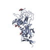 3zleSC S: Starting model for refinement C: citing same article ( |
|---|---|
| Similar structure data |


-Links
 PDBj
PDBj- Assembly
| Deposited unit | 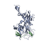
| |||||||||
|---|---|---|---|---|---|---|---|---|---|---|
| 1 |
| |||||||||
| Unit cell |
| |||||||||
| Components on special symmetry positions |
|
-Components
| #1: Protein | Mass: 42819.445 Da / Num. of mol.: 1 / Fragment: CONSERVED ECTOPLASMIC REGION, RESIDUES 97-388 Source method: isolated from a genetically manipulated source Source: (gene. exp.)  TRICHOPLUSIA NI (cabbage looper) / References: UniProt: B6K9M7 TRICHOPLUSIA NI (cabbage looper) / References: UniProt: B6K9M7 |
|---|---|
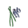
| #2: Protein/peptide | Mass: 4107.623 Da / Num. of mol.: 1 / Fragment: D3 REGION, RESIDUES 452-487 Source method: isolated from a genetically manipulated source Source: (gene. exp.)  |
| #3: Water | ChemComp-HOH /  Mass: 18.015 Da / Num. of mol.: 7 / Source method: isolated from a natural source / Formula: H2O Mass: 18.015 Da / Num. of mol.: 7 / Source method: isolated from a natural source / Formula: H2O |
| Has protein modification | Y |
| Sequence details | CORRECT SEQUENCE ONLY FOUND IN TOXODB, GENE TGME49_315730 |
-Experimental details
-Experiment
| Experiment | Method: X-RAY DIFFRACTION |
|---|
- Sample preparation
| Crystal | Density Matthews: 2.81 Å3/Da / Density % sol: 56.2 % / Description: NONE |
|---|---|
| Crystal grow | Details: 0.2 M MAGNESIUM CHLORIDE HEXAHYDRATE, 0.1 M HEPES PH 7.5, 25% PEG3350 |
-Data collection
| Diffraction | Mean temperature: 100 K |
|---|---|
| Diffraction source | Source: SYNCHROTRON / Site: SSRL  / Beamline: BL12-2 / Wavelength: 0.9795 / Beamline: BL12-2 / Wavelength: 0.9795 |
| Detector | Type: MARMOSAIC 325 mm CCD / Detector: CCD |
| Radiation | Protocol: SINGLE WAVELENGTH / Monochromatic (M) / Laue (L): M / Scattering type: x-ray |
| Radiation wavelength | Wavelength: 0.9795 Å / Relative weight: 1 |
| Reflection | Resolution: 3.1→85.96 Å / Num. obs: 9716 / % possible obs: 97.8 % / Observed criterion σ(I): 2 / Redundancy: 4 % / Rmerge(I) obs: 0.11 / Net I/σ(I): 8.2 |
| Reflection shell | Resolution: 3.1→3.27 Å / Redundancy: 3.7 % / Rmerge(I) obs: 0.31 / Mean I/σ(I) obs: 3.5 / % possible all: 95.8 |
- Processing
| Software |
| ||||||||||||||||||||||||||||||||||||||||||||||||||||||||||||||||||||||||||||||||||||||||||||||||||||||||||||||||||||||||||||||||||||||||||||||||||||||||||||||||||||||||||||||||||||||
|---|---|---|---|---|---|---|---|---|---|---|---|---|---|---|---|---|---|---|---|---|---|---|---|---|---|---|---|---|---|---|---|---|---|---|---|---|---|---|---|---|---|---|---|---|---|---|---|---|---|---|---|---|---|---|---|---|---|---|---|---|---|---|---|---|---|---|---|---|---|---|---|---|---|---|---|---|---|---|---|---|---|---|---|---|---|---|---|---|---|---|---|---|---|---|---|---|---|---|---|---|---|---|---|---|---|---|---|---|---|---|---|---|---|---|---|---|---|---|---|---|---|---|---|---|---|---|---|---|---|---|---|---|---|---|---|---|---|---|---|---|---|---|---|---|---|---|---|---|---|---|---|---|---|---|---|---|---|---|---|---|---|---|---|---|---|---|---|---|---|---|---|---|---|---|---|---|---|---|---|---|---|---|---|
| Refinement | Method to determine structure: MOLECULAR REPLACEMENT Starting model: PDB ENTRY 3ZLE CHAIN A Resolution: 3.1→85.96 Å / Cor.coef. Fo:Fc: 0.906 / Cor.coef. Fo:Fc free: 0.855 / SU B: 19.85 / SU ML: 0.353 / Cross valid method: THROUGHOUT / ESU R Free: 0.5 / Stereochemistry target values: MAXIMUM LIKELIHOOD / Details: HYDROGENS HAVE BEEN ADDED IN THE RIDING POSITIONS.
| ||||||||||||||||||||||||||||||||||||||||||||||||||||||||||||||||||||||||||||||||||||||||||||||||||||||||||||||||||||||||||||||||||||||||||||||||||||||||||||||||||||||||||||||||||||||
| Solvent computation | Ion probe radii: 0.8 Å / Shrinkage radii: 0.8 Å / VDW probe radii: 1.2 Å / Solvent model: MASK | ||||||||||||||||||||||||||||||||||||||||||||||||||||||||||||||||||||||||||||||||||||||||||||||||||||||||||||||||||||||||||||||||||||||||||||||||||||||||||||||||||||||||||||||||||||||
| Displacement parameters | Biso mean: 49.451 Å2
| ||||||||||||||||||||||||||||||||||||||||||||||||||||||||||||||||||||||||||||||||||||||||||||||||||||||||||||||||||||||||||||||||||||||||||||||||||||||||||||||||||||||||||||||||||||||
| Refinement step | Cycle: LAST / Resolution: 3.1→85.96 Å
| ||||||||||||||||||||||||||||||||||||||||||||||||||||||||||||||||||||||||||||||||||||||||||||||||||||||||||||||||||||||||||||||||||||||||||||||||||||||||||||||||||||||||||||||||||||||
| Refine LS restraints |
|