 Movie
Movie Controller

[English] 日本語
 Yorodumi
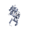
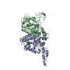
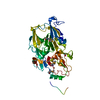
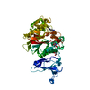
- PDB-3gis: Crystal Structure of Na-free Thrombin in Complex with Thrombomodulin -
+ Open data

- Basic information
| Entry | Database: PDB / ID: 3gis | ||||||
|---|---|---|---|---|---|---|---|
| Title | Crystal Structure of Na-free Thrombin in Complex with Thrombomodulin | ||||||
 Components Components |
| ||||||
 Keywords Keywords | BLOOD CLOTTING / protein-protein complex / coagulation / Acute phase / Blood coagulation / Cleavage on pair of basic residues / Disease mutation / Disulfide bond / Gamma-carboxyglutamic acid / Glycoprotein / Hydrolase / Kringle / Protease / Secreted / Serine protease / Zymogen / EGF-like domain / Hydroxylation / Membrane / Receptor / Thrombophilia / Transmembrane | ||||||
| Function / homology |  Function and homology information Function and homology informationblood coagulation, common pathway / apicolateral plasma membrane / serine-type endopeptidase complex / zymogen activation / vacuolar membrane / : / thrombospondin receptor activity / thrombin / thrombin-activated receptor signaling pathway / Defective factor XII causes hereditary angioedema ...blood coagulation, common pathway / apicolateral plasma membrane / serine-type endopeptidase complex / zymogen activation / vacuolar membrane / : / thrombospondin receptor activity / thrombin / thrombin-activated receptor signaling pathway / Defective factor XII causes hereditary angioedema / negative regulation of astrocyte differentiation / regulation of blood coagulation / neutrophil-mediated killing of gram-negative bacterium / positive regulation of phospholipase C-activating G protein-coupled receptor signaling pathway / Defective F8 cleavage by thrombin / ligand-gated ion channel signaling pathway / Platelet Aggregation (Plug Formation) / positive regulation of collagen biosynthetic process / negative regulation of platelet activation / negative regulation of blood coagulation / negative regulation of fibrinolysis / blood coagulation, fibrin clot formation / positive regulation of blood coagulation / Transport of gamma-carboxylated protein precursors from the endoplasmic reticulum to the Golgi apparatus / : / response to cAMP / Gamma-carboxylation of protein precursors / response to X-ray / Removal of aminoterminal propeptides from gamma-carboxylated proteins / regulation of cytosolic calcium ion concentration / fibrinolysis / : / negative regulation of proteolysis / negative regulation of cytokine production involved in inflammatory response / Regulation of Complement cascade / positive regulation of release of sequestered calcium ion into cytosol / acute-phase response / Cell surface interactions at the vascular wall / Peptide ligand-binding receptors / growth factor activity / positive regulation of receptor signaling pathway via JAK-STAT / female pregnancy / lipopolysaccharide binding / platelet activation / positive regulation of protein localization to nucleus / response to wounding / Golgi lumen / Regulation of Insulin-like Growth Factor (IGF) transport and uptake by Insulin-like Growth Factor Binding Proteins (IGFBPs) / positive regulation of reactive oxygen species metabolic process / blood coagulation / positive regulation of insulin secretion / transmembrane signaling receptor activity / regulation of cell shape / antimicrobial humoral immune response mediated by antimicrobial peptide / heparin binding / Thrombin signalling through proteinase activated receptors (PARs) / signaling receptor activity / positive regulation of cell growth / response to lipopolysaccharide / blood microparticle / G alpha (q) signalling events / positive regulation of phosphatidylinositol 3-kinase/protein kinase B signal transduction / cell surface receptor signaling pathway / endoplasmic reticulum lumen / receptor ligand activity / serine-type endopeptidase activity / external side of plasma membrane / signaling receptor binding / calcium ion binding / positive regulation of cell population proliferation / cell surface / proteolysis / : / extracellular exosome / extracellular region / plasma membrane Similarity search - Function | ||||||
| Biological species |  Homo sapiens (human) Homo sapiens (human) | ||||||
| Method |  X-RAY DIFFRACTION / SYNCHROTRON / MOLECULAR REPLACEMENT / molecular replacement / Resolution: 2.4 Å X-RAY DIFFRACTION / SYNCHROTRON / MOLECULAR REPLACEMENT / molecular replacement / Resolution: 2.4 Å | ||||||
 Authors Authors | Adams, T.E. / Huntington, J.A. | ||||||
 Citation Citation | Journal: J.Thromb.Haemost. / Year: 2009 Title: Molecular basis of thrombomodulin activation of slow thrombin Authors: Adams, T.E. / Li, W. / Huntington, J.A. | ||||||
| History |
|
- Structure visualization
| Structure viewer | Molecule: MolmilJmol/JSmol |
|---|
- Downloads & links
-Download
| PDBx/mmCIF format | 3gis.cif.gz | 267.3 KB | Display | PDBx/mmCIF format |
|---|---|---|---|---|
| PDB format | pdb3gis.ent.gz | 211.6 KB | Display | PDB format |
| PDBx/mmJSON format | 3gis.json.gz | Tree view | PDBx/mmJSON format | |
| Others |  Other downloads Other downloads |
-Validation report
| Arichive directory | https://data.pdbj.org/pub/pdb/validation_reports/gi/3gisftp://data.pdbj.org/pub/pdb/validation_reports/gi/3gis | HTTPS FTP |
|---|
-Related structure data

-Links
 PDBj
PDBj


- Assembly
| Deposited unit | 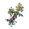
| ||||||||
|---|---|---|---|---|---|---|---|---|---|
| 1 | 
| ||||||||
| 2 | 
| ||||||||
| 3 | 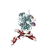
| ||||||||
| Unit cell |
|
-Components
-Protein/peptide , 1 types, 3 molecules ACE
| #1: Protein/peptide | Mass: 5641.175 Da / Num. of mol.: 3 / Fragment: Thrombin light-chain, UNP residues 315-363 Source method: isolated from a genetically manipulated source Source: (gene. exp.) Homo sapiens (human) / Gene: F2 / Plasmid: pET23 / Production host:  |
|---|
-Protein , 2 types, 6 molecules BDFXYZ
| #2: Protein | Mass: 29764.219 Da / Num. of mol.: 3 / Fragment: Thrombin heavy-chain, UNP residues 364-622 / Mutation: S195A Source method: isolated from a genetically manipulated source Source: (gene. exp.) Homo sapiens (human) / Gene: F2 / Plasmid: pET23 / Production host: #3: Protein | Mass: 12931.283 Da / Num. of mol.: 3 Fragment: Thrombomodulin EGF domains 4-5-6, UNP residues 363-483 Mutation: M388L,R456G,H457Q Source method: isolated from a genetically manipulated source Source: (gene. exp.) Homo sapiens (human) / Gene: THBD, THRM / Plasmid: pET39b / Production host: |
|---|
-Non-polymers , 3 types, 593 molecules 


| #4: Chemical | ChemComp-SO4 /  Mass: 96.063 Da / Num. of mol.: 15 / Source method: obtained synthetically / Formula: SO4 Mass: 96.063 Da / Num. of mol.: 15 / Source method: obtained synthetically / Formula: SO4#5: Chemical |  Mass: 40.078 Da / Num. of mol.: 3 / Source method: obtained synthetically / Formula: Ca Mass: 40.078 Da / Num. of mol.: 3 / Source method: obtained synthetically / Formula: Ca#6: Water | ChemComp-HOH / | Mass: 18.015 Da / Num. of mol.: 575 / Source method: isolated from a natural source / Formula: H2O |
|---|
-Details
| Has protein modification | Y |
|---|
-Experimental details
-Experiment
| Experiment | Method: X-RAY DIFFRACTION / Number of used crystals: 1 |
|---|
- Sample preparation
| Crystal | Density Matthews: 2.63 Å3/Da / Density % sol: 53.19 % |
|---|---|
| Crystal grow | Temperature: 294 K / Method: vapor diffusion / pH: 7 Details: 0.2M LiSO4, 22% PEG3350, pH7.0, vapor diffusion, temperature 294K, VAPOR DIFFUSION |
-Data collection
| Diffraction | Mean temperature: 100 K |
|---|---|
| Diffraction source | Source: SYNCHROTRON / Site: SRS  / Beamline: PX14.2 / Wavelength: 0.979 Å / Beamline: PX14.2 / Wavelength: 0.979 Å |
| Detector | Type: ADSC QUANTUM 4 / Detector: CCD / Date: May 20, 2007 / Details: mirrors |
| Radiation | Monochromator: Si 111 crystal / Protocol: SINGLE WAVELENGTH / Monochromatic (M) / Laue (L): M / Scattering type: x-ray |
| Radiation wavelength | Wavelength: 0.979 Å / Relative weight: 1 |
| Reflection | Resolution: 2.4→91.9 Å / Num. all: 60765 / Num. obs: 58334 / % possible obs: 96 % / Observed criterion σ(F): 0 / Observed criterion σ(I): 0 / Redundancy: 4.7 % / Rmerge(I) obs: 0.15 / Net I/σ(I): 8.9 |
| Reflection shell | Resolution: 2.4→2.53 Å / Redundancy: 3.2 % / Rmerge(I) obs: 0.339 / Mean I/σ(I) obs: 3.5 / % possible all: 95 |
-Phasing
| Phasing | Method: molecular replacement |
|---|
- Processing
| Software |
| ||||||||||||||||||||||||||||
|---|---|---|---|---|---|---|---|---|---|---|---|---|---|---|---|---|---|---|---|---|---|---|---|---|---|---|---|---|---|
| Refinement | Method to determine structure: MOLECULAR REPLACEMENT Starting model: PDB ENTRY 1JOU, 1DX5 Resolution: 2.4→40 Å / Occupancy max: 1 / Occupancy min: 1 / σ(F): 2 / Stereochemistry target values: Engh & Huber
| ||||||||||||||||||||||||||||
| Solvent computation | Bsol: 21.479 Å2 | ||||||||||||||||||||||||||||
| Displacement parameters | Biso max: 124.81 Å2 / Biso mean: 28.213 Å2 / Biso min: 2.83 Å2
| ||||||||||||||||||||||||||||
| Refinement step | Cycle: LAST / Resolution: 2.4→40 Å
| ||||||||||||||||||||||||||||
| Refine LS restraints |
| ||||||||||||||||||||||||||||
| Xplor file |
|
