 Movie
Movie Controller


+ Open data

- Basic information
| Entry | Database: PDB / ID: 1ey2 | ||||||
|---|---|---|---|---|---|---|---|
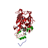
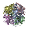
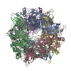
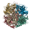
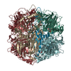
| Title | HUMAN HOMOGENTISATE DIOXYGENASE WITH FE(II) | ||||||
 Components Components | HOMOGENTISATE 1,2-DIOXYGENASE | ||||||
 Keywords Keywords | OXIDOREDUCTASE / jelly roll / beta sandwich | ||||||
| Function / homology |  Function and homology information Function and homology informationhomogentisate 1,2-dioxygenase / homogentisate 1,2-dioxygenase activity / Tyrosine catabolism / L-tyrosine catabolic process / L-phenylalanine catabolic process / extracellular exosome / metal ion binding / identical protein binding / cytosol Similarity search - Function | ||||||
| Biological species |  Homo sapiens (human) Homo sapiens (human) | ||||||
| Method |  X-RAY DIFFRACTION / Resolution: 2.3 Å X-RAY DIFFRACTION / Resolution: 2.3 Å | ||||||
 Authors Authors | Timm, D.E. / Titus, G.P. / Penalva, M.A. / Mueller, H.A. / de Cordoba, S.M. | ||||||
 Citation Citation | Journal: Nat.Struct.Biol. / Year: 2000 Title: Crystal structure of human homogentisate dioxygenase. Authors: Titus, G.P. / Mueller, H.A. / Burgner, J. / Rodriguez De Cordoba, S. / Penalva, M.A. / Timm, D.E. | ||||||
| History |
|
- Structure visualization
| Structure viewer | Molecule: MolmilJmol/JSmol |
|---|
- Downloads & links
-Download
| PDBx/mmCIF format | 1ey2.cif.gz | 103.5 KB | Display | PDBx/mmCIF format |
|---|---|---|---|---|
| PDB format | pdb1ey2.ent.gz | 77.6 KB | Display | PDB format |
| PDBx/mmJSON format | 1ey2.json.gz | Tree view | PDBx/mmJSON format | |
| Others |  Other downloads Other downloads |
-Validation report
| Arichive directory | https://data.pdbj.org/pub/pdb/validation_reports/ey/1ey2ftp://data.pdbj.org/pub/pdb/validation_reports/ey/1ey2 | HTTPS FTP |
|---|
-Related structure data


-Links
 PDBj
PDBj


- Assembly
| Deposited unit | 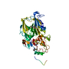
| ||||||||
|---|---|---|---|---|---|---|---|---|---|
| 1 | x 6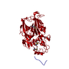
| ||||||||
| Unit cell |
| ||||||||
| Details | The biological assembly is a hexamer constructed from two-fold and three-fold crystallographic operations on the asymmetric unit. |
-Components
| #1: Protein | Mass: 53648.582 Da / Num. of mol.: 1 Source method: isolated from a genetically manipulated source Source: (gene. exp.) Homo sapiens (human) / Tissue: LIVER / Plasmid: PET19B / Production host:  |
|---|---|
| #2: Chemical | ChemComp-FE2 /   Mass: 55.845 Da / Num. of mol.: 1 / Source method: obtained synthetically / Formula: Fe Mass: 55.845 Da / Num. of mol.: 1 / Source method: obtained synthetically / Formula: Fe |
| #3: Water | ChemComp-HOH /  Mass: 18.015 Da / Num. of mol.: 232 / Source method: isolated from a natural source / Formula: H2O Mass: 18.015 Da / Num. of mol.: 232 / Source method: isolated from a natural source / Formula: H2O |
| Has protein modification | Y |
-Experimental details
-Experiment
| Experiment | Method: X-RAY DIFFRACTION / Number of used crystals: 1 |
|---|
- Sample preparation
| Crystal | Density Matthews: 3.19 Å3/Da / Density % sol: 61.39 % |
|---|---|
| Crystal grow | Temperature: 298 K / Method: vapor diffusion, sitting drop Details: Ammonium Sulfate, Imidazole, FeSO4, VAPOR DIFFUSION, SITTING DROP, temperature 298.0K |
| Crystal grow | *PLUS Method: unknown / PH range low: 7.4 / PH range high: 6.7 |
| Components of the solutions | *PLUS Conc.: 1.5-2.0 M / Common name: ammonium sulfate |
-Data collection
| Diffraction | Mean temperature: 100 K |
|---|---|
| Diffraction source | Source: ROTATING ANODE / Type: RIGAKU RU200 / Wavelength: 1.5418 |
| Detector | Type: RIGAKU RAXIS IIC / Detector: IMAGE PLATE / Date: Jun 25, 1999 |
| Radiation | Protocol: SINGLE WAVELENGTH / Monochromatic (M) / Laue (L): M / Scattering type: x-ray |
| Radiation wavelength | Wavelength: 1.5418 Å / Relative weight: 1 |
| Reflection | Resolution: 2.3→30 Å / Num. all: 31910 / Num. obs: 31495 / % possible obs: 98.7 % / Observed criterion σ(F): 0 / Observed criterion σ(I): 0 / Redundancy: 5.9 % / Biso Wilson estimate: 37.9 Å2 / Rmerge(I) obs: 0.083 / Net I/σ(I): 19.3 |
| Reflection shell | Resolution: 2.3→2.38 Å / Redundancy: 5.9 % / Rmerge(I) obs: 0.412 / Num. unique all: 2801 / % possible all: 89.9 |
| Reflection | *PLUS Num. measured all: 187375 |
| Reflection shell | *PLUS Highest resolution: 2.3 Å / % possible obs: 89.9 % / Mean I/σ(I) obs: 4.3 |
- Processing
| Software |
| |||||||||||||||||||||||||
|---|---|---|---|---|---|---|---|---|---|---|---|---|---|---|---|---|---|---|---|---|---|---|---|---|---|---|
| Refinement | Resolution: 2.3→30 Å / σ(F): 2 / σ(I): 0 / Stereochemistry target values: Engh & Huber
| |||||||||||||||||||||||||
| Refinement step | Cycle: LAST / Resolution: 2.3→30 Å
| |||||||||||||||||||||||||
| Refine LS restraints |
| |||||||||||||||||||||||||
| Software | *PLUS Name: X-PLOR / Version: 3.851 / Classification: refinement | |||||||||||||||||||||||||
| Refinement | *PLUS Highest resolution: 2.3 Å / Lowest resolution: 30 Å / σ(F): 2 | |||||||||||||||||||||||||
| Solvent computation | *PLUS | |||||||||||||||||||||||||
| Displacement parameters | *PLUS Biso mean: 37.9 Å2 |
