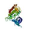
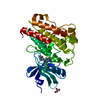
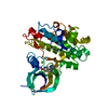
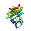
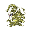
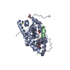
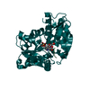
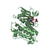
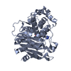
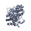
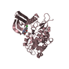
Entry Database : PDB / ID : 5qinTitle TGF-BETA RECEPTOR TYPE 2 KINASE DOMAIN IN COMPLEX WITH N- {4-[3-(6-METHOXYPYRIDIN-3-YL)-1H-PYRROLO[3,2-B]PYRIDIN-2- YL]PYRIDIN-2-YL}ACETAMIDE TGF-beta receptor type-2 Keywords / / / Function / homology Function Domain/homology Component
/ / / / / / / / / / / / / / / / / / / / / / / / / / / / / / / / / / / / / / / / / / / / / / / / / / / / / / / / / / / / / / / / / / / / / / / / / / / / / / / / / / / / / / / / / / / / / / / / / / / / / / / / / / / / / / / / / / / / / / / / / / / / / / / / / / / / / / / / / / / Biological species Homo sapiens (human)Method / / / / Resolution : 1.57 Å Authors Sheriff, S. Journal : ACS Med Chem Lett / Year : 2018Title : Discovery of 4-Azaindole Inhibitors of TGF beta RI as Immuno-oncology Agents.Authors: Zhang, Y. / Zhao, Y. / Tebben, A.J. / Sheriff, S. / Ruzanov, M. / Fereshteh, M.P. / Fan, Y. / Lippy, J. / Swanson, J. / Ho, C.P. / Wautlet, B.S. / Rose, A. / Parrish, K. / Yang, Z. / ... Authors : Zhang, Y. / Zhao, Y. / Tebben, A.J. / Sheriff, S. / Ruzanov, M. / Fereshteh, M.P. / Fan, Y. / Lippy, J. / Swanson, J. / Ho, C.P. / Wautlet, B.S. / Rose, A. / Parrish, K. / Yang, Z. / Donnell, A.F. / Zhang, L. / Fink, B.E. / Vite, G.D. / Augustine-Rauch, K. / Fargnoli, J. / Borzilleri, R.M. History Deposition Aug 5, 2018 Deposition site / Processing site Revision 1.0 Oct 31, 2018 Provider / Type Revision 1.1 Dec 26, 2018 Group / Database references / Category / citation_authorItem _citation.journal_abbrev / _citation.journal_volume ... _citation.journal_abbrev / _citation.journal_volume / _citation.page_first / _citation.page_last / _citation.pdbx_database_id_PubMed / _citation.title / _citation_author.name Revision 1.2 May 12, 2021 Group / Structure summaryCategory / pdbx_struct_conn_angle / struct_connItem _pdbx_deposit_group.group_type / _pdbx_struct_conn_angle.ptnr1_auth_seq_id ... _pdbx_deposit_group.group_type / _pdbx_struct_conn_angle.ptnr1_auth_seq_id / _pdbx_struct_conn_angle.ptnr1_symmetry / _pdbx_struct_conn_angle.ptnr3_auth_seq_id / _pdbx_struct_conn_angle.ptnr3_symmetry / _pdbx_struct_conn_angle.value / _struct_conn.pdbx_dist_value / _struct_conn.ptnr2_auth_seq_id / _struct_conn.ptnr2_symmetry Revision 1.3 Mar 6, 2024 Group / Database references / Category / chem_comp_bond / database_2Item / _database_2.pdbx_database_accessionRevision 1.4 Feb 18, 2026 Group / Structure summaryCategory / pdbx_initial_refinement_model
Show all Show less
 Movie
Movie Controller

 Yorodumi
 Open data
 Basic information
 Components
 Keywords
 Function and homology information
 Homo sapiens (human)
 X-RAY DIFFRACTION /
  Authors
 Citation
 Structure visualization
 Downloads & links
 Other downloads
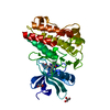


 PDBj
PDBj


 Assembly

 SPODOPTERA FRUGIPERDA (fall armyworm)
 Mass: 359.381 Da / Num. of mol.: 1 / Source method: obtained synthetically / Formula: C20H17N5O2
 Mass: 24.305 Da / Num. of mol.: 1 / Source method: obtained synthetically / Formula: Mg
 Mass: 92.094 Da / Num. of mol.: 1 / Source method: obtained synthetically / Formula: C3H8O3
 Mass: 18.015 Da / Num. of mol.: 218 / Source method: isolated from a natural source / Formula: H2O
 Sample preparation
 / Beamline: 17-ID / Wavelength: 1 / Wavelength: 1 Å
 Processing